
There are no products in your shopping cart.
THE Stuff Interloping and Overwhelming Everything: Examples and Ideas for Remediation in Our Immediate Environments
Wed, 08/12/2015 - 3:24pm

INTRODUCTION to THE Stuff
I've created content extensively in 2015 on "THE Stuff", as I coined the term to make it non-specific.
THE Stuff interloping in 'everything', and changing everything once you learn about it, and how it 'operates'.
Since we're all learning about this, and are clearly outsiders from the 'insiders' who do research and don't tell the general public until they want to in whatever version of the truth they disclose, what we know changes all the time too. So I'm editing this on March 22, 2016 -- over 5,000 people had read this topic so far. I hope they come back and re-visit, reinforce what we had here before by being reminded, and seeing what is NEW.
RE-editing again about two months later, May 23, 2016, to make this more apparent about where I encountered this information, and what the consensus is among people I know online who have also studied with 'the teacher', Steve Beddingfield. I don't know a person who got into "Steve's Images" on Facebook who wasn't perplexed by Steve's behavior if you talked to them privately. Or collectively, in other groups.
I was in the group for eight months before Steve made time to talk to me individually by phone, and he gave me a lot of time over the next few months to learn the information and include it at Lumigrate. I don't know a person who I've communicated with privately, the ones perplexed by the overall about Steve, who is NOT APPRECIATIVE of the opportunity they had when finding him and his information to learn. Many kept a distance, lurking in his Facebook group to keep up on any new developments. But there were a lot of 'falling outs', and things that just don't add up to everything being 'clean' surrounding who he is, what his motives are, etc.
"I'm just sayin'.....". If you go to his group, or befriend him on Facebook or wherever, buyer beware, but also please see the positives I have included, and that literally everyone felt they learned and grew. That was then, this is now, and I have no idea how things are going there now.
I chose to keep giving input in the group while he was replacing the 'leaders' who were to 'divorce' in April of 2015, leaving a gap in competence with advisement in my opinon. If I'm having content on Lumigrate, I want people to be able to be properly supported if they're needing help. So I'm going to say this loud and clear at this point in the topic, adding in up near the top so it cannot be missed. Contact me if you find this information and want to learn more about it. There are too many wonky things that occur in Steve's Images to ONLY go there to learn. I'm not saying don't go there, just don't go there without having someone who understands the history and wonky-ness also guiding you.
There was another 'housecleaning' this year, this time May (2016) which apparently stemmed from Steve's learning of a group of us who'd moved on and were discussing many other things related to strategies to reverse symptoms in addition to using reishi and fenbendazole potentially, which is what his protocol is 'based in'. Considering the length and depth of interaction I had with Steve, it would seem appropriate to contact me privately if he learned of something that he thought was inappropriate on my part, or offensive.
But that is not how it was handled, what occurred was unfriending, removing from Steve's Images group, so now if anyone going there from Lumigrate is in the group, I'm not able to see the threads and shore up anything that's not said accurately. He has had one administrator who took a lead early on after finding the group, allegedly after finding Lumigrate's coverage of it, who has had enough time now that she's providing accurate advise consistently.
But this type of unprofessional, unkind, immature behavior is NOT what I want YOUsers of Lumigrate to be exposed to! It's up to YOU, go there if you wish, but I would prefer people not get into the 'spin cycle' that everyone I've mentored so far through his group have had go on. Just trying to figure out why he doesn't answer questions directly, makes it more confusing than it needs to be -- there's not a person who doesn't wonder 'what is up with this' and thing 'something isn't adding up'. Having said that, I remind of what I'd said, above: Everyone who's learning the information has been appreciative of it, when they move on, sometimes in disgust with the behaviors and quandries.
This is not an indictment of anyone helping Steve, but of Steve for not having people really knowledgeable about things before admins are allowed to give advise. This latest admin was giving better advise with each week in the group than I would have been giving with that amount of time. This is not the easiest information to learn enough to teach others and guide others with questions. However, there appears to have been lack of wisdom of what she found out by being included in another group and relating that to Steve. He somehow had the impression that I had the group he learned of -- I'm not even an administrator nor a very robust contributor. That or he knew the truth and lied and told someone to defame my character and knew otherwise. A previous administrator, who was running his group when I tuned in, has had similar behaviors in the last six months that are just not things you encounter when dealing with professionals who have their heads and ethics straight. Hence my concern for YOUsers of Lumigrate.
I had verbalized recently in Steve's Images that I was about to drop back out of participation so much, so this is not an upsetting thing to me for my sake, aside from I would have liked to have kept up on new developments. However, when Steve said something to the extend of why I'd put 'crap' like info about chemtrails on my website, I would answer that. When he stated that the 'shooter at Sandy Hook' was an example of someone with certain issues, I suggested saying 'alleged shooter', because of the numbers of people who believe it's a false flag event. This has to make one wonder if Steve is part of disinformation dissemination on the part of a program or not.
So find me on Facebook (Mardy Ross, very easy to find), go to the About tab and look at other contact information that's updated there, or the blog tab, the pinned blog about Mardy Poppins is also updated. (The Contact Us I have been able to change once in the past but now cannot see how to update, so it's not necessarily correct, I hope in the future to solve that and maybe the contact us will be correct, but at the time of this edit it is not.)
10,865 reads on August 16, 2017 with a very slight re-edit while looking over the thread to share in Crystal Tripp's group about Dis-Ease Solutions (which currently is starting to discuss The Stuff, the strands, the bizarre and 'difficult' which can become divisive, so let us hope with her leadership and expertise first hand from her perspective (in light of the phone time we've had to discuss this aspect of illness and reversing symptoms), it's a good place to go.
IF you're finding this thread, I'll suggest you connect with me and I'll help facilitate getting you into the group. My area code is 970, and the prefix is 462 and the last for are 8662. email is mardy dot poppins at yahoo dot com .... and my Facebook -- MESSAGE ME if you are putting in a friend request if I don't respond right away. If you're wanting in that group reach me somehow.
You can, of course, request to join but Crystal is very busy and sometimes those things are not as high priority to those busy as getting other things done such as serving the people who have already subscribed and are ... making you busy! It's great, people finding the information and products .... and we all just have to make our adjustments to this new life of working together to reverse all this mess going on. Without overwhelming ourselves and crashing health-wise.
One of the people who found this information (and/or other topic in this forum, which is similar) , allegedly, via 'Google' was a fast learner and extremely motivated to get solutions to her health problems as they had drastically affected her life.
(Notice my insertion and editing in August 2017 of the word 'allegedly', because as time has gone on and the discussions have gone on about what his group is really doing, and who's been in there, I just continue to be suspicious that things are often not what people say they are in terms of these subjects that are being disclosed in a slow drip way to the public, and using Facebook is clearly one very potent place to disclose to people. There are good groups out there funding and there are the groups out there massively funded to do what's been happening for 'ever', leading people to believe what we have been lead to believe and it's really almost impossible to be sure who's on which team.)
Because she epitomizes what the Lumigrate YOU! model is about, and in honor of this being the anniversary month for Lumigrate's being here for SEVEN YEARS (live on the Internet since late March 2009, after 1-2 years of research, planning, and development / creation) providing 'the truth as best I can find to provide', I'm going to prominently put a few of the things she provides to her Facebook followers here on Lumigrate. With my gratitude and thanks.
This is the picture that I think was her #1 contribution and started a great thread of information in Steve's Images Facebook group:
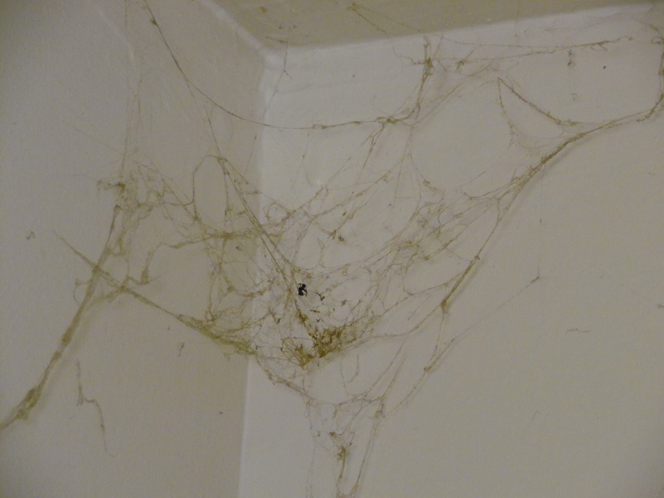
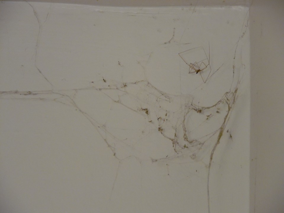
Here's another one which shows a different type of spider's body -- you can see why we thought these were 'spider webs' when in face the spiders are utilizing and being symbiotic with the slime mold / myxomycetes.
I'd realized in 2015 that THE Stuff was what was on and in the computers, and sought out having mine cleaned professionally but didn't take the time to photograph it. She did, thankfully, and this is one of her photographs I felt was going to do a great job of bringing this information 'home' to everyone searching online about complex chronic illness on the Internet .... most are using or have used computers such as these (and hopefully still are since screen size really assists in better seeing and therefore understanding this information).
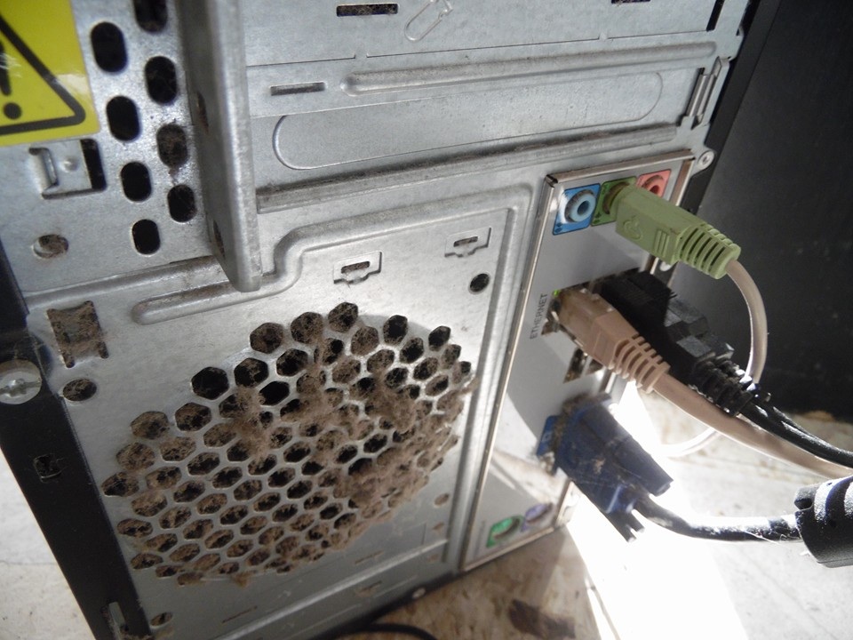
Her words: "This is not dust. Parasitic slime mold called labyrinthulomycetes."
Photo credit to the afore-mentioned woman who, as I said above, brought joy to me in terms of the ripple effect of learning involved with someone finding Lumigrate's information and directing them to a learning place in Facebook where they became a significant contributor. May she find the joy of returning wellness level, return to job searching (protected by the anonymity here with photo credits, knowing I'm giving credit where credit (and thanks) is due.
She had her learning curve and then had to figure out how to change her exposures. She taught those around her in her personal life who could grasp and further the ball, so to speak, and then made a plan to move. Once moved, since she didn't want to mix things up with those who owned the housing, she blasted her Facebook with what she wanted people to know.
I'm therefore adding in, below, information in her honor about " slime mold " / " myxomycetes " , because that's what she brought to the forefront of the discussions (in Steve's Images Facebook group) and furthered our understanding of what we have in our environment and inside of us causing us to be unhealthy.
SLIME MOLD / MYXOMYCETES
To get started, here's one resource about it, which includes photographic images of how visually intriguing they are: myxomycetes.net/
AND please take the time to go and read what is at this link at Wikipedia.
en.wikipedia.org/wiki/Dictyostelium_discoideum
It turns out that the image of the life cycle that's at the above link is able to be used if given credit, so I'm going to give the 'picture that's worth a thousand words' here WITH credit to the source, clearly. In the blockquote area below, you'll find the image plus the section of the topic at Wikipedia about the 'symbiosis' between it and bacterial prey.
"Farming"[edit]
A 2011 report in Nature published findings that demonstrated a "primitive farming behaviour" in D. discoideum colonies.[18][19] Described as a "symbiosis" between D. discoideum and bacterial prey, about one-third of wild-collected D. discoideum colonies engaged in the "husbandry" of the bacteria when the bacteria were included within the slime mold fruiting bodies.[19] The incorporation of the bacteria into the fruiting bodies allows the "seeding" of the food source at the location of the spore dispersal, which is particularly valuable if the new location is low in food resources.[19] Colonies produced from the "farming" spores typically also show the same behavior when sporulating. This incorporation has a cost associated with it: Those colonies that do not consume all of the prey bacteria produce smaller spores that cannot disperse as widely. In addition, much less benefit exists for bacteria-containing spores that land in a food-rich region. This balance of the costs and benefits of the behavior may contribute to the fact that a minority of D. discoideum colonies engage in this practice.[18][19]
D. discoideum is known for eating Gram-positive, as well as Gram-negative bacteria, but some of the phagocytized bacteria, including some human pathogens,[20] are able to live in the amoebae and exit without killing the cell. When they enter the cell, where they reside, and when they leave the cell are not known. The research is not yet conclusive but it is possible to draw a general lifecycle of D. discoideum adapted for farmer clones to better understand this symbiotic process.
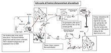 Lifecycle of farmer D. discoideum
In the picture, one can see the different stages. First, in the starvation stage, bacteria are enclosed within D. discoideum,[20] after entry into amoebae, in a phagosome the fusion with lysosomes is blocked and these unmatured phagosomes are surrounded by host cell organelles such as mitochondria, vesicles, and a multilayer membrane derived from the rough endoplasmic reticulum (RER) of amoebae. The role of the RER in the intracellular infection is not known, but the RER is not required as a source of proteins for the bacteria.[21] The bacteria reside within these phagosomes during the aggregation and the multicellular development stages. The amoebae preserve their individuality and each amoeba has its own bacterium. During the culmination stage, when the spores are produced, the bacteria pass from the cell to the sorus with the help of a cytoskeletal structure that prevents host cell destruction.[22] Some results suggest the bacteria exploit the exocytosis without killing the cell.[22] Free-living amoebae seem to play a crucial role for persistence and dispersal of some pathogens in the environment. Transient association with amoebae has been reported for a number of different bacteria, including Legionella pneumophila, many Mycobacterium species, Francisella tubarensis, and Escherichia coli, among others.[21] Agriculture seems to play a crucial role for pathogens' survival, as they can live and replicate inside D. discoideum, making husbandry. Nature’s report has made an important advance in the knowledge of amoebic behavior, and the famous Spanish phrase translated as “you are more stupid than an amoeba” is losing the sense because amoebae are an excellent example of social behavior with an amazing coordination and sense of sacrifice for the benefit of the species.

Presuming you are convinced by the information about it presented here and upon your further searching/researching, this topic is intended to help YOU 'light the path' to your wellness level improvement or maintenance.
I suggest a person seeing this do a quick looking over of the photographs included, below, and the section headings so you know the overview and overall. I'm editing in mid November, the summary provided today in the Facebook group where I've learned about THE Stuff, and continue to keep abreast of things. There is a separate topic at Lumigrate about our being disclosed to -- things being revealed to us in many ways that is different information than what we'd been lead to believe in the past -- about history -- about science -- about health. The mainstream media and social media are tools that those who want information to get to us utilize. And then there are independent, free people who are truly not affiliated and being coerced or paid as motivation for doing things as they do.
It's never easy to 'follow the money', or 'follow the coersion', but we must all do our best with that. I am pleased to say I am not being encumbered by anyone -- there is no advertising at Lumigrate, I've not had any financial or legal problems that someone bailed me out of with strings attached, and I make abundant information totally free on Lumigrate. You don't even have to give an email signup, as a matter of fact that's been filled up for a year so if someone volunteers to sign up, it's not registering. If someone wishes to have my assistance and contacts me, then it might be that I can help them or refer them on to others who can. That's where I get my support, and then I support Lumigrate solely. (The About tab is where you can find how to contact me).
By mid November there had been a lot of disclosure steps going on in the mainstream and more people were coming to the Facebook group, and the leader had either entered a new phase of what he's allowed to say or he's gotten his way of telling people things improved based on his figuring things out more as he went along. As one person goes, we all go, really.... we are all connected more than people realize I think. So here's the summary from mid November, 2015 from Steve Beddingfield:
Super heated water exits volcanic chambers,
rocks there are decomposed by the water,
water carries nutrients upward to hydrothermal vents,
sediment is created,
life forms live off these nutrients -- sulphur, iron, copper, cobalt, manganese,
Life forms such as bacteria, fungi, protit's, dinoflagellates, plankton, all thrive on these nutrients, no light exists, they have adapted to chemosynthetic processes, now that they have arrived here on continents, it is we who must now adapt to them. This adaption is very difficult to figure out, but here in this group, we are doing a great job of figuring this out. This group is on the front lines and the battle is not being lost.
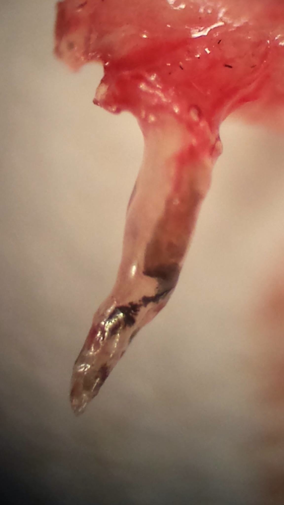
Worm in picture is able to penetrate bones, muscle tissue ..... It softens the bones by destroying components within them, calcium is depleted with the worms, which search out and use phosphorus. 60X magnification, taken from a skin lesion.
I might add that how it has arrived on continents has taken place across history's timeline, it's just happened MORE frequently and in higher quantities due to the way we've had man-made and nature-made weather events and other things disrupting them. They also are fed by other things than the minerals talked about, above, we've put phosphates and things from petroleum into the environment in massive amounts.
Picture a spiral, it was a spiral of things over a long period of time. Disorder of wellness has always occurred, it's just that we've seen the age of onset of things shift to younger and more dramatic in recent generations. In my generation, I was perhaps one of the more affected kids in my class growing up in the mountains outside of Denver, Colorado (USA) but I was thought to be 'healthy'.
I had learning problems that were dismissed, physical symptoms that were dismissed -- even when they'd take me to a doctor for something, looking back on what the symptoms were and knowing what I have learned since, they were missing the boat so much that I can see why my parents didn't take me in unless something seemed really serious. You can see the effects of the disorder of my system in pictures, and you can see it in others in my class. But if one compares a photo of my class photograph to one of similar aged children today, it's clear things are much worse two generations (or fifty years) later.
Yes, it's fifty years since I headed to public school. I'm appreciative of all my hard experiences AND to have something to present here to YOUsers that might help them see that it's not natural for us to have learning problems, it's very normal now since so many brains are unwell because so many people are affected by THE Stuff in a significant enough way.
This Stuff is a situation that takes a long time to explain, AND then it takes a long time for the receiver to absorb it -- work with it, replace what we thought was reality and what to do and not do, and then do differently. It has affected everything on Earth. It's causing massive problems and your health and life depend on your learning this. So get an idea by looking over this topic and seeing 'the path' you can go on to find solutions -- that don't cost a lot of money, but take time and therefore energy to learn. Decide what your priorities are. Think about your overall life and where for a short time you can spend less or no time on something and use that time to learn what's here.
The path you'll go on ends up with a lot of pieces that play together, but the root of the matter is that marine bacteria that originally came up in vents from below the surface of the Earth/ocean floor, always through time, have overall gotten more "stirred up" by big weather events in recent years and decades, such as Hurricane Katrina as one example. It uses things for fuel that are bioaccumulating more in the Earth's environment due to all the things we've done in modern times without understanding or perhaps caring enough about long-term consequences.
I'd say overall, it was likely both. If you're a person who won't make the time and use the energy to look enough at this information, you'll get the results from that which will be your feedback. It's as simple as that. To make something simplified but also provide enough information is something which takes me and others helping me enormous amounts of time, so I've done my part. I created a trough, you've gotten to it however you have, so it's up to you to drink enough ... or not. It's THAT simple.
Beyond Bacteria
Snails, Slugs, and Semi-slugs: A Parasitic Disease in Paradise
Posted on April 3, 2009 by

CDC plays a vital role supporting state health departments, particularly with management of rare or lesser-known pathogens. Recently, CDC’s Division of Parasitic Diseases (DPD) was contacted by the Hawaii Department of Health (HI DOH) for advice regarding three cases of presumedAngiostrongylus cantonensis (AC) infection. AC, commonly called the rat lungworm, is a parasitic worm and the most common infectious cause of eosinophilic (a type of white cell) meningitis in humans worldwide.
AC has an interesting life cycle. Infected rodents carry the adult worm and pass immature worms in their feces. Mollusks (i.e., snails, slugs, or semi-slugs) become infected by ingesting immature worms in the rat feces. Humans become infected by ingesting raw or undercooked mollusks (these guys can be tiny enough to hide on a nickel; – check it out[PDF, 1 page]! infected with the worms or contaminated raw produce. Transmission might also occur through ingestion of raw or undercooked freshwater shrimps/prawns, crabs or frogs. In humans, AC causes eosinophilic meningitis, the symptoms of which can include headache, stiff neck, nausea, vomiting, low-grade fever, fatigue, and abnormal skin sensations (e.g., tingling or pain). In most cases, the symptoms disappear in weeks to months and most patients recover completely, although rare cases of blindness, paralysis, and death have been reported.
 A picture from differential interference contrast microscopy showing A. cantonensis. This larva was obtained from a P. martensi slug collected in Hawaii. Infective, third-stage larvae measure 0.425 mm – 0.523 mm in length
Armed with this information, a team of scientists from USDA (HI), HI DOH and DPD began testing mollusks collected near the case-patients’ homes in Hilo Puna. We used morphologic and molecular techniques to test and document infection in slug samples sent from Hawaii. HI DOH has determined that many patients live in communities outside of the municipal water/sewage system and consume home-grown produce. The DPD molecular diagnostic parasitology lab, which I lead, continues to collaborate on testing of environmental samples and the development of methods to evaluate the effectiveness of interventions to eliminate infected rodents and mollusks. In this new phase of the collaboration, a real-time PCR assay developed in-house at DPD to detect A. cantonensis is being used to analyze mollusk samples collected in Hawaii. Future efforts will involve the transferring of this real-time PCR test and other molecular methods to Hawaii so testing can be performed in-state.
Since initial contact with Hawaii, four more cases of presumed AC infection have been reported. In response, we are helping to educate the public in Hawaii on how to prevent the infection. Information has also been developed for the healthcare community in Hawaii because many physicians may not consider AC when evaluating patients with eosinophilic meningitis. AC can be prevented by avoiding the consumption of raw/undercooked snails, slugs, freshwater shrimp/prawns, crabs and frogs; by washing raw produce thoroughly prior to eating; and by wearing gloves and washing hands after handling mollusks.
Posted on April 3, 2009 by
--- from the following link (please go and see the comments, it's been moderated by Alex and was very informative. AND, I might point out, from 2009.
From 2012, June, and a mainstream media source: http://discovermagazine.com/2012/jun/03-hidden-epidemic-tapeworms-in-the-brain
Hidden Epidemic: Tapeworms Living Inside People's Brains
Parasitic worms leave millions of victims paralyzed, epileptic, or worse. So why isn’t anyone mobilizing to eradicate them?
By Carl Zimmer|Tuesday, May 15, 2012

A human brain overrun with cysts from Taenia solium, a tapeworm that normally inhabits the muscles of pigs.
Courtesy of Theodore E. Nash , M.D.
Theodore Nash sees only a few dozen patients a year in his clinic at the National Institutes of Health in Bethesda, Maryland. That’s pretty small as medical practices go, but what his patients lack in number they make up for in the intensity of their symptoms. Some fall into comas. Some are paralyzed down one side of their body. Others can’t walk a straight line. Still others come to Nash partially blind, or with so much fluid in their brain that they need shunts implanted to relieve the pressure. Some lose the ability to speak; many fall into violent seizures.
Underneath this panoply of symptoms is the same cause, captured in the MRI scans that Nash takes of his patients’ brains. Each brain contains one or more whitish blobs. You might guess that these are tumors. But Nash knows the blobs are not made of the patient’s own cells. They are tapeworms. Aliens.
A blob in the brain is not the image most people have when someone mentions tapeworms. These parasitic worms are best known in their adult stage, when they live in people’s intestines and their ribbon-shaped bodies can grow as long as 21 feet. But that’s just one stage in the animal’s life cycle. Before they become adults, tapeworms spend time as larvae in large cysts. And those cysts can end up in people’s brains, causing a disease known as neurocysticercosis.
“Nobody knows exactly how many people there are with it in the United States,” says Nash, who is the chief of the Gastrointestinal Parasites Section at NIH. His best estimate is 1,500 to 2,000. Worldwide, the numbers are vastly higher, though estimates on a global scale are even harder to make because neurocysticercosis is most common in poor places that lack good public-health systems. “Minimally there are 5 million cases of epilepsy from neurocysticercosis,” Nash says.
He puts a heavy emphasis on minimally. Even in developed nations, figuring out just how many people have the illness is difficult because it is easy to mistake the effects of a tapeworm for a variety of brain disorders. The clearest proof is the ghostly image of a cyst in a brain scan, along with the presence of antibodies against tapeworms.
The closer scientists look at the epidemiology of the disease, the worse it becomes. Nash and other neurocysticercosis experts have been traveling through Latin America with CT scanners and blood tests to survey populations. In one study in Peru, researchers found 37 percent of people showed signs of having been infected at some point. Earlier this spring, Nash and colleagues published a review of the scientific literature and concluded that somewhere between 11 million and 29 million people have neurocysticercosis in Latin America alone. Tapeworms are also common in other regions of the world, such as Africa and Asia. “Neurocysticercosis is a very important disease worldwide,” Nash says.
Cyst Attack
The alarming illness occurs when tapeworm larvae lose their way. Normally,Taenia solium has a life cycle that takes it from pigs to humans and back to pigs again. Adult tapeworms, living in the intestines of humans, produce up to 50,000 eggs apiece. The eggs are shed in the infected person’s feces. Pigs swallow these eggs accidentally as they rummage for food on the ground. When the parasite eggs reach a pig’s stomach, larvae hatch and burrow their way into the animal’s bloodstream. Eventually they end up lodged in small blood vessels, typically in the animal’s muscles. There they form cysts and wait until their host is eaten by a human. (Pork has to be undercooked for the tapeworms to complete their journey.)
But sometimes tapeworms take a wrong turn. Instead of going into a pig, the eggs end up in a human. This can occur if someone shedding tapeworm eggs contaminates food that other people then eat. When the egg hatches, the confused larva does not develop into an adult in the human’s intestines. Instead, it acts as it would inside a pig. It burrows into the person’s bloodstream and gets swept through the body. Often those parasites end up in the brain, where they form cysts.
The tapeworm larvae often get stuck in ventricles, or fluid-filled cavities, in the brain, sprouting grapelike extensions. In this way the worm actively cloaks itself from immune cells. Protected and well fed, its cysts can thrive there for years.
As a tapeworm cyst grows, it may push against a region of the brain and disrupt its function. It may get stuck in a passageway, damming the flow of cerebrospinal fluid. This impasse can cause hydrocephalus, or water on the brain, along with dangerously high pressure. A resulting brain hernia can result in stupor, coma, or death.
If a tapeworm cyst doesn’t cause big troubles, it may go unnoticed for its entire life. Eventually a tapeworm cyst that can’t move on to its adult stage will die; this signals the host’s immune system, eliciting a powerful attack and bringing its covert deception to an end. In many cases, the immune cells swiftly annihilate the revealed cyst, but often damage occurs. The immune system’s attack on the cyst can cause the surrounding brain tissue to swell with inflammation. For reasons unknown, a calcified cyst can keep triggering these immune reactions for years after the parasite’s death.
Although any cyst in a susceptible area of the brain can cause seizures, those lodged near regions that issue commands to muscles can trigger violent convulsions. One of Nash’s patients suffered from tapeworm cysts that twisted around his brain stem. After the tapeworms died, the inflammation that followed was so severe it put the man in a coma.
“Thirty or 40 years ago, these patients just died. Surgeons would go in and see this mess and couldn’t do much,” Nash says. Fortunately, the situation is improving. Even his comatose patient woke up and, after a few years of off-and-on treatment, completely recovered. “Now the guy is doing quite well.”
Breaking the Cycle
A great step forward came in the mid-1980s when praziquantel, the first drug able to kill tapeworm larvae in the brain, became widely available. But praziquantel proved too effective. It not only kills tapeworms but also triggers an immune reaction that causes brain swelling. “Paradoxically, we produce the disease we want to treat,” Nash says.
Over the years Nash and others refined the treatment by combining praziquantel with other drugs that tamp down the immune system. It is far from a perfect solution, though. Sometimes the immune system still overreacts, requiring years of care for seizures and other symptoms. And immune-suppressant drugs like steroids have side effects of their own.
The hunt for better drugs to fight neurocysticercosis is not an easy process. The best way to test potential medicines on tapeworms is to get living cysts out of infected pigs. Nash and his colleagues recently set up a lab in Peru, where infected pigs are abundant, to do just that.
Although finding a better cure is important, Nash is more interested in preventing tapeworms from getting into human brains in the first place by breaking their life cycle. A favored strategy is identifying people who have adult tapeworms in their bodies and giving them drugs to kill the parasites. It is also possible to vaccinate pigs so that they destroy tapeworm eggs as soon as they ingest them.
None of this is rocket science—which makes Nash all the more frustrated that so little is being done. “I see this as a disease that can be treated and prevented,” he says. But there are precious few resources available for treatment and little recognition of the problem. “All of this seems to be very feasible, but nobody wants to do anything about it.”
From 2013, and the 'biggie C word that gets people's attention', at the Science 2 point 0 website:
http://www.science20.com/catarina_amorim/new_mechanism_disco...
New Mechanism Discovery: How A Parasite Causes Cancer


About 200 million people across 75 of the poorest countries in the world are now infected by the blood parasite Schistosoma haematobium (S. haematobium). The infection causes severe urogenital disease, but also causes bladder cancer in a number of patients and why this occurs is not clear.
Now a group of Portuguese scientists believe they have the answer – their research shows how the parasite’s eggs can make human bladder cells behave as cancerous cells. And the key to that – according to the first author of the work Mónica Botelho– are catechol oestrogens, a molecule derived from estrogen (the sex hormone) that was found by the researchers in the eggs and is known to be highly carcinogenic (causes cancer).
Now a group of Portuguese scientists believe they have the answer – their research shows how the parasite’s eggs can make human bladder cells behave as cancerous cells. And the key to that – according to the first author of the work Mónica Botelho– are catechol oestrogens, a molecule derived from estrogen (the sex hormone) that was found by the researchers in the eggs and is known to be highly carcinogenic (causes cancer).
The research, a collaboration of the CECA/ICETA from the University of Porto, the National Institute of Health in Porto, Portugal and the George Washington University, US could be a first step towards one day be able to identify S. haematobium infected patients at risk of bladder cancer or even prevent the cancer by targeting catechol-oestrogens. Schistosomiasis is also associated to fertility problems and the newfound molecules might hold the key to also understand this.
Schistosomiasis, despite the numbers infected, remains a neglected tropical disease that affects the world’s poorest with a socioeconomically impact in the developing world only second to malaria. The disease is transmitted to humans by freshwater snails from contaminated waters, with the worms entering our blood stream to release eggs that become embedded in the bladder wall where they cause chronic inflammation and, in some patients, lead to bladder cancer.
How common is this carcinoma among parasite-infected patients is difficult to knowbecause the most affected countries are also the world’s poorest with scarce or even non-existing disease recording.
Nevertheless, in Egypt, disappearance of S. haematobium saw the type of tumors associated with the infection going from being almost 80% of all diagnosed bladder cancers, to less than 27 %, suggesting that the infection leads to a significant number of cancer cases.
S. haematobia life cycle
Botelho and colleagues have been investigating this relationship for many years, and shown already that extract from the adult worm could make animal cells acquire cancer-like characteristics and even form tumors if injected into mice with no immune system, further proving the parasite-cancer link.
Following the finding that Schistosomiasis’ patients had higher than normal levels of oestrogens Botelho and colleagues also discovered new estrogenic molecules released by S. haematobium. These molecules down-regulate estrogen receptors effectively blocking the host’s oestrogens (that act through these receptors).This, as Botelho explains “was an important clue because we know that oestrogen receptors are reduced when cancer becomes more invasive”.
The new molecules were later identified as a combination of DNA and catechol oestrogen-quinones (a derivate of estrogen). Catechol estrogens have been linked to several types of cancer, including breast and prostate cancer, suggesting that the new molecules could be the link between schistosomiasis and bladder cancer.
The research now published follows these results looking at the effect of S. haematobium eggs (the parasite stage associated with the cancer development) on normal human bladder cells. For that Botelho and colleagues exposed the cells to extract from the eggs and found that treated cells, when compared with normal control cells, divided much more, died much less and showed signs of oxidative stress.
Uncontrolled cell division and resistance to die are hallmark characteristics of cancer, and oxidative stress is known to be implicated in cancer formation.
To confirm that these changes were linked to cancer Botelho and colleagues next looked for DNA lesions. If DNA - the cell’s “instruction book” - becomes damaged and is not properly repaired, it will start giving wrong “instructions”, which can lead to the abnormal behavior typical of cancer (uncontrolled cell multiplication, “immortality”, etc.). And in fact, exposure to parasite’s eggs was linked to a visible increase in DNA lesions in the cells. The eggs were confirmed to contain the same new estrogenic molecules found in adult worms.

S. haematobia eggs embedded in the bladder wall (Image from CDC Public Health Image Library)
Based on the new data, Botelho and colleagues are now proposing a mechanism for the Schistosomiasis-bladder cancer connection.
As Botelho explains, ”What we think happens is that the parasite releases oestrogen molecules into the host. These are metabolized into catechol-oestrogen quinones, which are known to have high affinity for DNA and as result form estrogen-DNA adducts that can lead to the bladder cancer."
In fact, adducts , defined as pieces of DNA covalently bonded to a cancer-causing chemical, are known to interfere with normal cell division increasing the chance of DNA mutations and, consequently, of cancer. The carcinogenic effect of this estrogen–DNA adduct could then explain the link between S. haematobium infection and the carcinoma.
Botelho and colleague’s work have several implications – the possibility of using the new identified estrogenic molecules as biomarkers for bladder cancer in Schistosomiasis patients, or even as therapy targets for a start.
This is important because although at the moment it is suggested that there are about 4 cases of cancer for 100 000 Schistosomiasis-infected individuals what does not seem much, but we must remember that 200 millions people are believed to be infected, and even those numbers, like the Egypt case suggests, are probably a gross underestimation. The reality is that carcinoma of the urinary bladder is the most common malignancy in the Middle East and parts of Africa where schistosomiasis is a major problem.
Not only that but, and despite the existence of a cheap and effective drug, the disease (which is asymptomatic until very late) seems to be increasing and spreading. This is probably due to the large numbers of economical migrants from developing countries, as well as the wars in these areas of the globe, that create large displacements of people.
Another interesting potential implication for Botelho’s results is the possibility that the new identified estrogenic molecules could have a role in other cancers associated with infection and estrogenic changes, such as cholangiocarcinoma, a liver cancer linked to an infection by a parasitic liver fluke.
A question remains though - why does the parasite produce estrogenic molecules? An option, according to the researchers, could be that uses them to reduce the density of the bladder wall (a known effect of reduced estrogen receptors). After all S. haematobium eggs must cross the bladder mucosa to be excreted in order to survive and continue its life cycle. Another possibility is that the parasite is manipulating the host’s hormonal environment to improve its own living conditions.
Part of Botelho’s future work will be looking at the effect of the new estrogenic molecules on the parasite life cycle.
Citation: Botelho, M.C., et al. Tumour-like phenotypes in urothelial cells after exposure to antigens from eggs of Schistosoma hae- matobium: An oestrogen–DNA adducts mediated pathway? Int. J. Parasitol. (2013), http://dx.doi.org/10.1016/j.ijpara.2012.10.023
TO GET STARTED WITH SOME VISUALS AND OVERVIEW "At the Top":
In trying to provide, up front, a picture that will do the best job for most people to see what THE Stuff is, and how it gets inside of us, into our intestines where it over time --- all along -- has been creating it's environment for growing and using what else is down there, making the gut's biosphere it's very own, I'm starting with the photo that, from ALL The photos I've seen in the last year about THE Stuff, is the most impactful for starting out. Used on other topics at Lumigrate, it's showing us the way ingestion gets THE Stuff into us, and makes us aware about our food quality._jpg%20.jpg)
I went to 'smell the roses' that bloom one last time in the Grand Valley of Western Colorado, so this is a photo I took in October 2015. See the very thin strand going away from the camera, from the rose to the big bush?
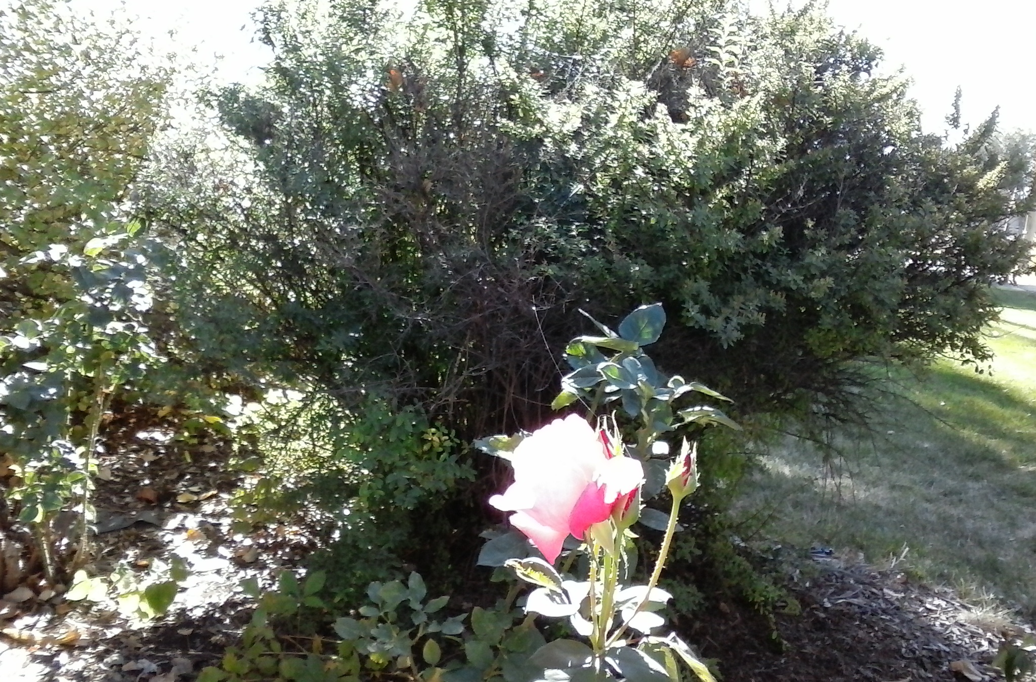
There were tons of other strands visible, but you have to catch them glinting in the sun. Look at your surroundings where YOU ARE. "Notice", pay attention. "Attend". Look inside your home, as "THE Stuff" ends up INSIDE too, they can be very long going from a piece of furniture to the ceiling or a wall, or just dangling from the ceiling if they've latched onto something there. They are easily spotted in corners or spaces where they've caught on something .... windows, or where the paper comes out of a printer -- use the light right to spot and you'll make this learning experience easier or faster.
I found one that had grown a 'feather'-like part hovering over a chair that I'd just been sitting in the day before -- that was back in August. There was a shadow from the 'feather' on the seat cushion of the chair, which was in the sun. My brain at the time was set for hornets and wasps as there'd been a lot of those in the area and I wasn't wanting to get 'stung'. Well, THE Stuff will sting you too, it's an acid-like burn you might feel. To me when I grabbed globs of The stuff, either as a 'feather' or a blob on the butt of the dog who'd been in a ditch (and then you guessed it, clean the dog...), it felt to my fingers like it 'buzzed'. What causes that sensation .....
Around that time, summer 2015, with a sudden wind storm, from the west -- 'blowy' some would call it -- I felt something wrap around my arms as I held them in front of me. I'd seen these starting in 2013 and didn't know what they were, did not understand the connection to the dying foliage, to my symptoms, to my beloved's symptoms (in that case a cat, who had to be euthanized). WHAT TO DO ABOUT IT has EVERYTHING TO DO WITH CLEANING --- out our intestines (but that has to be done with much learning and deciding by YOU what's best for YOU) and systems overall ... and everything we're exposed through. So, much about cleaning follows.
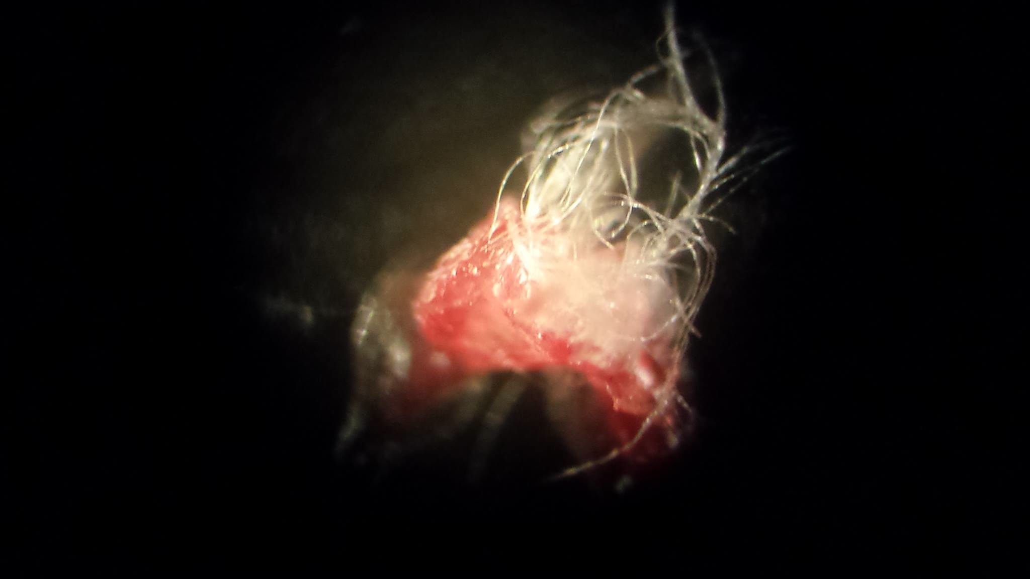
(The white strands are THE Stuff. Arcobacter bacteria, a type of giant sulful bacteria / GSB, Steve Beddingield said. This was only "discovered" by scientists, as Steve related, about a year ago -- I'd change that word to "revealed" because I think Steve knows that much is known by research insiders that is not released to the public UNTIL IT IS ALLOWED TO BE by those who control what's out here for people to learn.
It's a 'mat maker', he goes on to tell me, and the organisms feed off the mat and live in the mat and become symbiotic with the mat, and that explains the snail. The mat on the bottom of the ocean floor ---- or the mat coating your intestines (small intestines). Stop here. Process this, I suggest. Then go on. Take notes if that will benefit, draw things out on a piece of paper and 'connect' with the information. LEARN THIS. DO differently because of what you LEARN. Or not. Up to you.
Wikipedia has stated that arcobacter can be pathogentic to mammals: humans and other animals. The snail's DNA became symbiotic with the mat because of the GSB (Giant Sulphur Bacteria) (arcobacter in this case) -- the white strands would be somewhat the mat-forming bacteria, and others would be thioploca beggiatoa, Steve suspect. There are not many types of GSB disclosed to the public and independent researchers such as Steve naturally are at a disadvantage, not only with funding but with what is available to them. You'll find information about Steve's crowd source funding if you continue on learning the information and taking the links I provide, below. Please consider sacrificing something you'd do that's not a necessity in your day, week, month, year, and do so. Even if it's having one less drink out at your favorite drink place, that money will provide him a great amount compared with what he allegedly has to work from financially at this time.
This pickle also had an extra surprise for the consumer, as you see. This is a commercially prepared, name brand pickle. A mother following Steve Beddingfield's work and protocol had her teen daughter see this on her second bite into the pickle. The daughter had been not "buying in", had been 'resisting' or 'pushing back' with what the mother attempted to provide to her to treat her gut, as ideally one has to treat the whole system for THE Stuff. That changed with this pickle; thanks to the anonymous mom who approved use of this picture. ©2015 Lumigrate)
Example of what you'll see relative to what gets on you and your clothing (and your pets and in your house and in your yard, on your vegetables and ...... ) 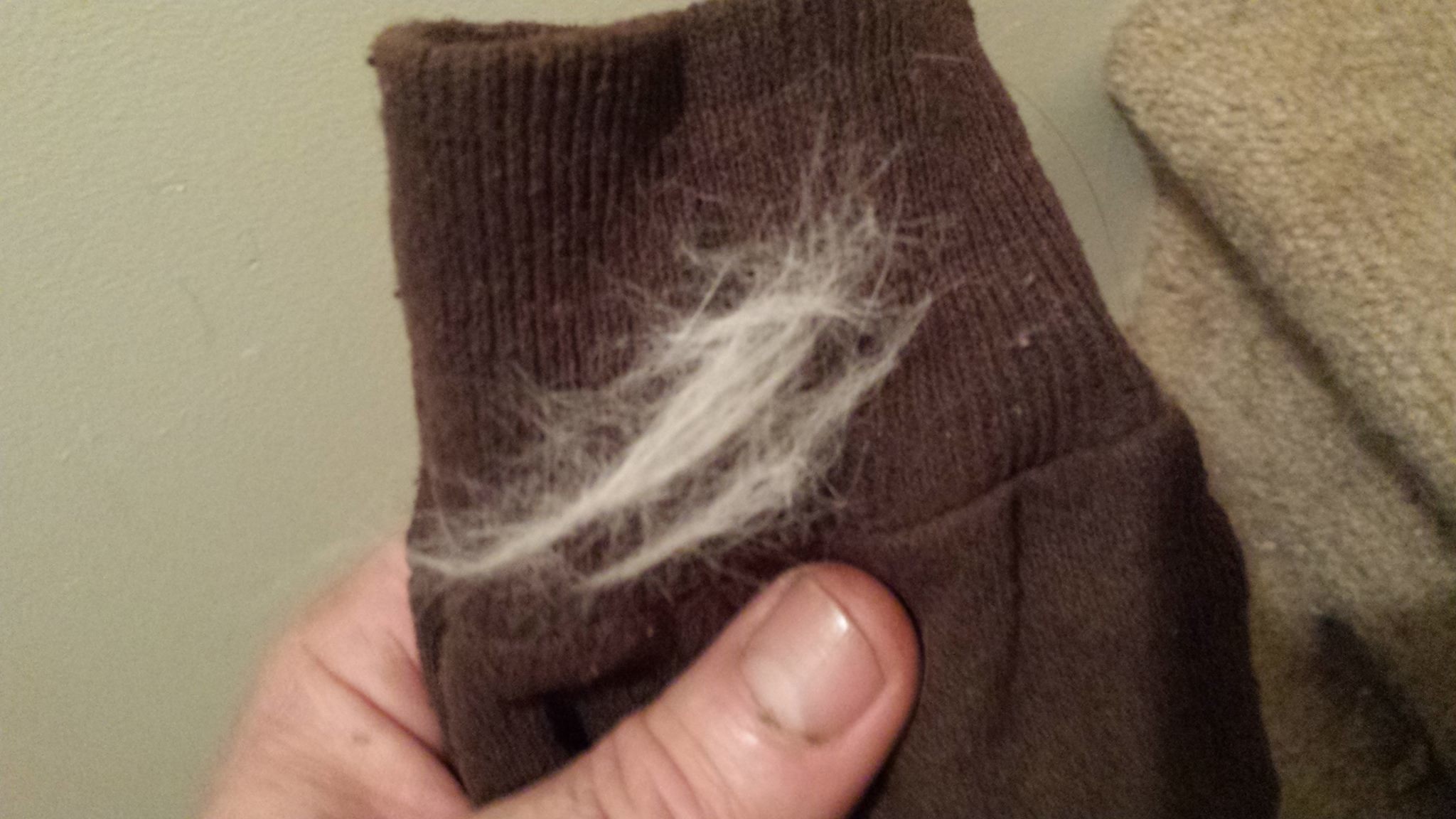
Both photos © 2015 Steve Beddingfield "Tubules" "This was on the floor of my buddy's hot rod garage. It was on some 90-weight gear oil, which is rich in sulfur, which makes it smell like what is referred to as 'sour crude'. The Gulf of Mexico always has sour crude." You'll also see this photo again in this topic, later. BELOW, tubules microscopically photographed by Steve, from marine bacteria.
THE BASICS
These bacteria, originally 'marine bacteria' become 'terrestrial bacteria' when they are transported up into the atmosphere in the clouds and then come down onto the land (soil), icepack, snowpack, and watery surfaces of Earth. This 'stuff' interplays with what's in the environment (as I said, above, feeding on things we're having more of .... phosphates, petroleum / plastics .... radiation .....).
That's the first piece of the puzzle to 'nail down', think of it as building a 100 piece puzzle and how you'd start with the good corner piece and get started with that, perhaps. This is a major situation which has always been what's behind illnesses in people and other living things, but it's been percolating faster and moreso as the interplay of things which allow it to grow versus things that would keep it in check have changed in our modern, industrial era and 'times'. Depending where on Earth you are, it's either about to go 'off the chain', or it already is, everywhere you've been seeing chemtrailing it certainly has been because that's a reason for the project, clearly kept secreted as long as possible for obvious reasons (more about that, below, for those who might not understand why at this point.)
MY POINT OF REFERENCE, MY REQUEST FOR SUPPORT
I think I'm in an interesting postion with the combination of my having encountered the researcher Steve Beddingfield in late summer of 2014 and at Christmastime realized the validity of his work thanks to his then accomplis, who I call 'PosterGal' on Lumigrate. Learning with the 1-2,000 people in his group, dozens of whom have been very active in sharing and learning together, in addition to time 1:1 with Steve via telephone, now almost daily as things appear to be 'going off the chain' with the heat and humidity of summertime 2015 in full bloom, so to speak, I've been literally scrambling to get things understood by me, take samples, photographs, and ask for guidance, then bring what I feel is right to bring to YOUsers at Lumigrate.
Due to my physical position in the Grand Valley of central, western Colorado, about as 'inland' as one can be, we're perhaps a bit 'behind the curve' compared to areas nearer the bodies of water where these marine bacteria originally were living and multiplying. I have a good 'baseline' of understanding since I've lived in western Colorado since late 2003, and on the Front Range of Colorado from the lower mountains outside of Denver, to Fort Collins, then Denver, back to the mountains, back to Denver, then on to Colorado Springs before migrating west.
It's inverse in the 'banana belt' of Colorado --- the winters are mild and you can comfortably walk outside most of the days in the winter but in the summers the heat is limiting for those of us without perfect wellness to withstand extremes of temperature. I adjusted within two years, to being aware of how I needed to find a way to be active and keep moving in the summertime whereas in the Front Range area it was the winter my health would slide backwards. Overall my wellness did better in western Colorado, as the story goes with Lumigrate, hence when I figured out things that I thought others could benefit from, I'd put it on the website I'd created in 2007-9 in order to share the good news I was learning.
Since many of my old friends are on the Front Range, I'm interested in what they'll encounter as they learn about THE Stuff. I believe the overall Colorado area, and other surrounding states, are 'better off' with 'THE Stuff' than many other places and we will be welcoming continued growth into the area, as people will transplant to somewhere it's relatively easier to survive with THE Stuff and the realities it is bringing with it in terms of how we live. How we clean, how our yards look, what kind of home we live in, our clothing materials and what it covers or does not cover, how we launder/clean our clothing, our bodies and hair -- how we wear our hair on our heads, faces, bodies ... how we grow our foods, our water sources, EVERYTHING is changing due to THE Stuff. It will be in your best interest to learn this information.
So far, the responses I see by those who have the capacity to take in new information with an open mind (a prerequisite), when I present in person to people about THE Stuff who were unaware of it, typically is a 'that connects dots for me' reaction, OR even at a more subtle level of 'this resonates for me'. Some will listen and just be holding onto it to consider. I've noticed in one case of someone I think important to have be aware in this community about The Stuff, from one time to the other they appear to not recall that I've even told them about it before!
THE SPIRITUAL ASPECT
So this is a good place to remind everyone about the spiritual aspect of integrative medicine; not everyone has a life path where they're going to get the information everyone's been progressing along with. They are with their own realities on Earth. Don't worry about them, perhaps hold space for their wellness as they go on with their reality and where it leads. We have to be realistic about the many who will be injured because they're just not processing correctly, who will die of unforseen causes, who will die from diseases in increased numbers beyond what we've ever experienced before, and illnesses which often are excruciating to experience.
Steve Beddingfield experienced that, coming close to death when he was 55 years old, the age I am today. I have as well, knowing at age 40 that if I didn't turn things around, I didn't want to live very long. By 45 I had turned things around and had a mini celebration for my birthday that year because I had secretly known and feared that I'd be dead by 50 if I didn't invest my time, energy, and money -- which I thankfully had one good year of income and my father offered to help in the second year so I could continue on with my treatments and testings (things I now know were unnecessary, thanks to what you can DIY via what Steve Beddingfield figured out and shares with others how to do).
So here I am, having created and continued to nurture and grow Lumigrate.com thanks to my father's having set aside some of his money for me via his will, and due to his cousin who had done the same which occurred just as I had wished I had money to create a website to further the successful education 'forum' I was doing 'live and in person' in 2007. I mention this because I think it's important for people using Lumigrate to understand, via transparency, why the resource is here. Your support of my work is necessary, no matter what your relationship is with me. Thanks to all who do what they can and do what they do. Even if that's simply getting the word out to others, that's support. And it goes from there.
Steve Beddingfield repeatedly cites God/Source as being a guide for his prayers to heal himself and his part of the bargain was to give the information to others. In a way, he reminds me of a brotherless version of "My Name is Earl", the popular television show about the man who is reformed and redeeming all his wrongs, an average guy who is clearly very intelligent and enterprising, going through a lot because of the people surrounding him physically and in relationships. My progress with information is repeatedly hindered because of the interruptions and sidetracking that happens. So the information here 'is what it is', it's my best attempts at this time to get it from Steve, use it myself, and then provide it for YOUsers at Lumigrate.
MY UNIQUE POSITION of KNOWLEDGE LEVEL AND LOCATION, PLUS WORKING WITH CONSUMERS AND BUSINESS PEOPLE ALLOW ME TO SEE HOW TO IMPLEMENT THE INFO ABOUT "THE Stuff"
I was the first move of All Pro Moving of Grand Junction, in early January 2004, and they still tell tales of their trek over the Rockies to retrieve and move my household possessions, aside from what I'd brought in my sedan in two trips in December, thinking I was coming to work for three weeks as an occupational therapist, initially, then feeling I was just supposed to be here for whatever reason Source/God had in mind. At that time, I did not realize our weather was being geoengineered --- I'd known of Colorado's famous 'cloud seeding' in the 1960s but then all the talk about that had gone away in my circles.
Slammed with a snowstorm, I prepared the entryway to my new home using my snow brush and scraper for my car so they could 'hit it fast with all men on deck' to unload the van that Sunday morning. The Denver Broncos were on that day, in playoffs, so their workers seemed to 'come out of the woodwork' and before you knew it, my stuff was inside and they were gone. This is, to some extent, an analogy for what I'm seeing occur with 'THE Stuff' -- massive numbers of what we think are old fashioned 'bugs' of whatever kind appear overnight and leave evidence behind, things that had just never happened before.
THE PEP TALK
May we have the serenity, as the infamous prayer says. Made famous because of it's use in mainstream addiction 'recovery' circles, programs, businesses, I believe it provides a good backdrop for 'this stuff'. There's a lot we're going to have to accept, some of it we can change and some of it we cannot. And we'll have to have to figure out which is which. And then have enough 'left in us' to take action on what we wish to be proactive about. And to realize there have been other ways of looking at addiction recovery with a 20% rate of relapse, whereas conventional, mainstream had 80% relapse, 20% do well.... Reinforcing the need to study the models out there, decide what kind of experts you're going to follow and learn from, ask for help from, etc.

I've also covered a bit about THE Disclosure, which I believe we are in. I felt we were being disclosed to about UFOs about five years ago based on what was being shown on television, including the ABC Comedy show "The Neighbors" which was aired right after their smash hit on Wednesday evenings, Modern Family. I felt The Colbert Report was used in December 2013 relative to geoengineering, and am curious about the timing of Dave Letterman leaving and Mr. Colbert taking his seat in the near future.
Relative specifically to THE Stuff interloping in everything and affecting everything, which this topic is intended to give an overview of what it is and what to do to reduce exposures, we'll be hearing about this stuff from someone besides Steve Beddingfield, and me, and the other few thousand of his followers who presumably have an influence in passing information along to others in a 'ripple effect'.
And I think we'll be hearing from mainstream about this very soon, based upon what I've seen in the new season on History Channel of the show Ancient Aliens, which moved from the less-establishment-approved "H2" at some point. The information they've hinted at or laid the groundwork to reveal more about in future episodes appears to me to be substantiating things that THE Teacher of THE Stuff (so far), Steve Beddingfield, has done his best to present after figuring things out as best he can with his brain, tools, experience, and dedication.
He is one of millions who has suffered the devastation of health and finances when a person has a complicated case of chronic, environmentally-caused, illness. He feels he was giving the ability to figure out what this is about and present it, and so he takes that responsibility seriously. As do I. I am, today, mid 50s, the age Steve was when his health collapsed and he started trying to figure THE Stuff out. I was in my late 20s when my life-long symptoms that were kept 'in the background' one day were suddenly fore-ground: I went to bed one night like any other night and went to get up the next morning to go to work and simply could not get my eyes to focus, the strength and energy to get up were just not there.
And so it began. In 2007 in Grand Junction, Colorado, where I'd lived since 2004, I'd have a live education group about reversing symptoms and dealing with complex chronic illness, where The YOU! Model, the cornerstone of Lumigrate's information and my method of teaching consumer empowerment, was first utilized.
![]() EMPTY YOU! Model, YOU are the center, fill it in -- who do you want around YOU, providing services, products, etc.? What happens with this information you're looking at right now -- up to YOU. Below's just one way it might shape up ....
EMPTY YOU! Model, YOU are the center, fill it in -- who do you want around YOU, providing services, products, etc.? What happens with this information you're looking at right now -- up to YOU. Below's just one way it might shape up ....
.jpg)
Mentioning "Ancient Aliens" will likely not find YOU neutral about that topic -- it might seem a negative thing that I'd mention that program, it might be a positive. I've seen it work both ways. Let me further clarify that I'm not a regular viewer of the show, as it has felt, like so much put out by mainstream, to have an agenda and could have what I'd call propaganda, mind control, etc. I don't totally avoid movies and television and popular music, I try to 'enjoy them' but keep in mind to be thinking about what they're 'doing' with what they're providing. What's their 'deal' that's not being told to us outright.....
I'd heard that Ancient Aliens was going to have something on a new episode that was related to the cloning that Steve's photographs show, via Steve telling people in his group. So I found what channel it was on. JUST in time for the blitz they ran over July 4 weekend in pre-paring people for the NEW season. They were basically showing the key older episodes with perhaps segments in them that, it appeared to me, chain together quite interestingly into what is a disclosure. Right or wrong, truth or not, YOU will have to decide. But The System which owns The Media is Disclosing ..... It's Happening...
Just today Steve and I both had time to go over the pictures I'd taken from his group page on Facebook to use on Lumigrate, and it was clear that he's simply stating what he thinks things are, and then learning more and what he'll figure later is different than sooner. Can't we all commiserate with that? However, when he types things in his group page on Facebook it sounded or was interpreted by me to be more 'factual'.
So I hope people read this and understand that I'm providing what I have. And this is what I have. Take it. Leave it. Up to YOU! Decide for yourself. Find him on Facebook or his group and see more for yourself. It took me six months to get 'into it' and that was at Christmas/New Years, so I've immersed in this all year and the more time goes on, the more comfortable I am with that this is at least 'on the right track' and will be helpful for any one who learns of it. So, I provide. That's all I can do (aside from providing individualized learning with people, naturally).
He doesn't add in all the thoughts and caveats he adds in when he's on the phone.... In his southern, North Carolina accent, notably not 'backwoods' sounding and with very good grammar and manners, I notice, much is being lost in translation to the written, readable with him. I hope he very soon is recorded by someone very good at interviewing on information like this. It will very much help his information reach more people to hear him speak about this. It seemed to me from talking with him he's extremely sincere, serious but with a sense of humor, grounded well, and wanting to help the overall society. In big ways. I kept having to say 'trees, not forest, now, please'. He obliged. This takes a lot of time, so if the information I provide is 'blog style', please understand I'm melding my desire for high standards with the necessity to get this information here for YOUsers to utilize, ASAP.
I do believe he's authentic, and has a good grasp of this information he has been researching and presenting to others. A mechanic by background, he became ill seven years ago, 2008; he was in his mid 50s, the age I am now. He thinks of himself as much a "researcher" presently and going forward as I do an Internet - based teacher and information provider and consultant, but what we discussed today was about how we needed to use the knowledge from our younger adulthood and middle adulthood to help people figure out how to clean the insides of their cars and maintain them differently in light of this information we know and people will be turned onto. Yards. Gardens. Homes.
My background as an occupational therapist, teaching people things about activities of daily living having to do with their surgery or illness, which insurance-based medicine was paying for, provided me with a better vision of how to be helping people in their homes and businesses, perhaps. I hope our collaboration which I bring with this topic is going to be of value to you. If you can reciprocate, I hope you do. If you can help forward the information, I hope you do.
Steve had outward skin lesions as a primary symptom, which helped him to identify as having 'Morgellons Disease", and he'd found at a nearby mobile home on or near his property at the worst of his wellness, a child's toy microscope; so he started trying to figure things out. One of his first photos is of what appears to be some type of insect (cloned insect) that has no eyes, so he presumes it was something that came from the ocean and was spewed up into the clouds and to his home, where he'd found the 'thing'. The STUFF he figured out, started with The Thing. Steve even enrolled in college to try to get the skills he needed to learn and do better faster and found out that is not what college is about. He says he likes to drink two beers a day, that would be a goal to be able to have a couple of beers AND the things he needs to eat AND do his research.
He has a small Airstream trailer and a pickup truck. He thinks about going around and seeing what's going on with other places in the country since we all talk about it in his group. His dog just died, he said he buried it not very deep and a long ways from the house.
THE Stuff was all over the ground at the grave site within days. He's seen a grave where a cat was buried not far from a house have The Stuff have the intelligence somehow to know how to replicate and move all the way along the ground to where the bedroom was in the house, and up the walls into the house and to the bed. Steve surmises the bacteria had gotten DNA from the cat, to explain what he saw that day.
He's posted this about cats:
From feces from Bernie the 21 year old cat: ©2015 Steve Beddingfield
But he has seen similar coming from dog vomiting up something similar in appearance after being treated with fenbendazole. 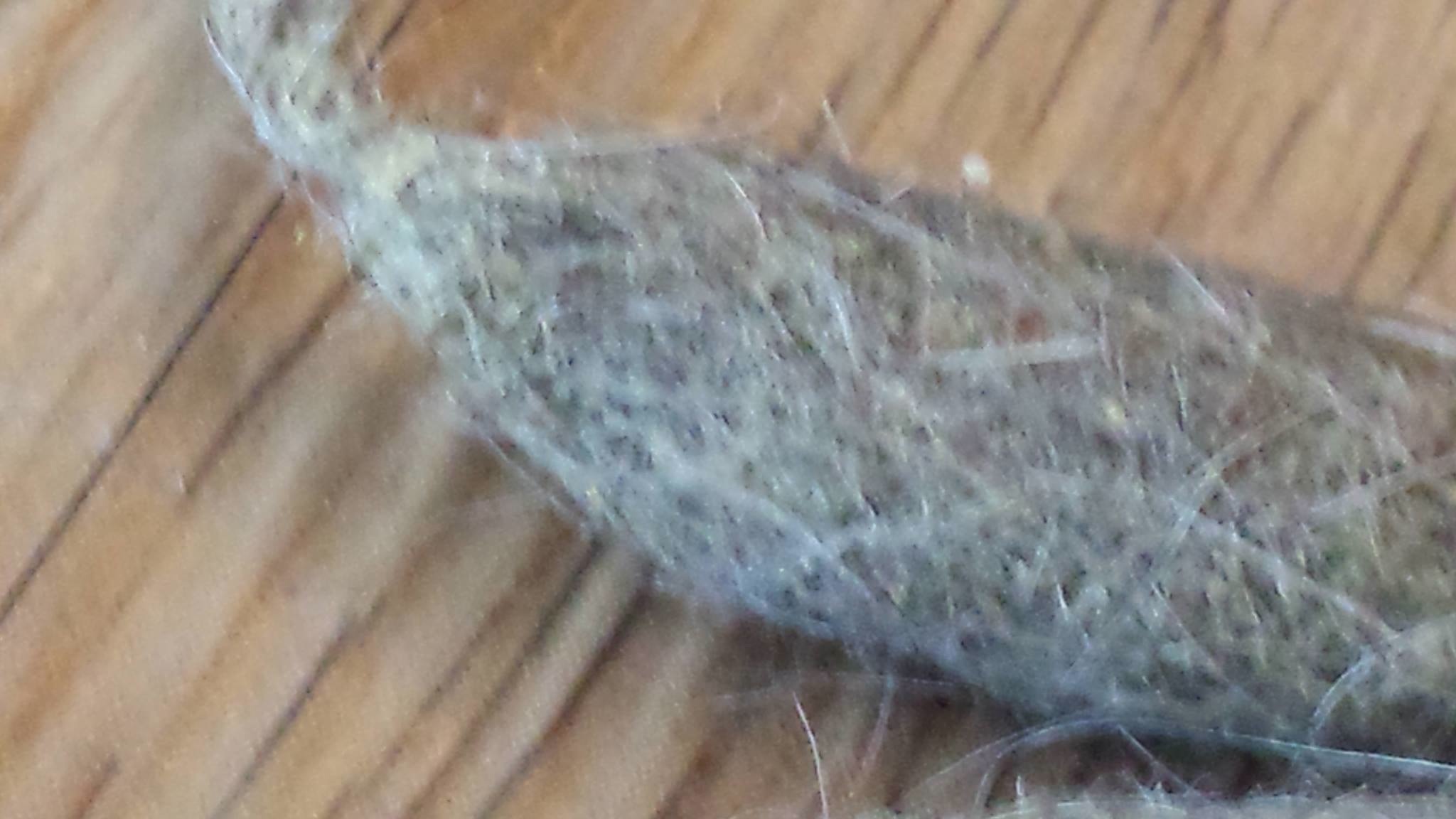
Pets (Cats and Dogs as Focus of This Introductory/Basic Topic)
The comment at the long, complex topic (in this same forum) about Steve's work, that is specific about pets / animals, for those wanting more, is at this link: www.lumigrate.com/forum/steve-his-images-his-protocol-and-facebook-group-community-and-other-resources-chronic-illness#comment-2814
For Cats: (From September 9, 2015, from Steve Beddingfield's group consultation)
©
 ©2015 Mardy Ross SpoildeyCat as Baby Cat (Had THE Stuff, had to be euthanized before age 4 after having episodes of seizure-like behavior where she'd think what she saw had to be driven away, couldn't tell reality from what occurred in an episode. Sensory integration symptoms, autism symptoms. She grew into 'my greatest 10# teacher' I called her. RIP. Teach in your absence, through your story.)
Piperizine cat dewormer internally. For the coat use fenbendazole spray: mix one ounce of fenbendazole liquid suspension (for goats) to 25 ounces of water, spray twice daily. (Or we've discussed before, you spray it on a cloth or some sort of wiper tool and wipe the animal).
After working with many cats, kittens, it's clear to me just how important is to treat them early on, they suffer greatly and (he believes, surmises) the bacteria picks up the genes of Bartonella and babesia from the cats, using lateral gene transfer, then donates these pathogens to the cats owner. Hell begins for the owner. Never allow cats to rest on one's bed, especially if not healthy. Piperizine monthly and fenben wipes daily or weekly will ensure safety for the owner and will make for a beautiful cat.
For Dogs
See link, above
Does THAT get your attention? Maybe YOU need to have a beer or two or whatEVER you 'do' to help 'digest' things that are stressful. Wine. Camomile tea, any kind of tea. Coffee. But --- is it with a lot of THE Stuff in/on it? Is the manufacturer or producer, grower 'aware', and doing what can be done to have the product as safe as can be prior to your having it 'in your hands'. Some would maybe do some more deep breaths. Whatever it is, take care of YOU and do your best to learn this IF you're seeing it's value.
Becoming ill in recent years since Facebook, as Steve did, has so many great people on it communing about common interests is a whole different ballgame than before the Facebook era. Naturally, the Internet generally makes it a whole new era too, compared to when my health had two major downhill events in the late 1980s and mid 1990s. He'd not really seemed to know a lot about that though, it's almost as though he'd not learned all the things about diet and leaky gut and things that 'we all have learned by now' if we have kids with autism symptoms or ourselves have that or more fatigue or neurodegenerative and 'fibromyaligia' symptoms. Point being, because of connections and Facebook, here we are with YOU now having this information.
The photo I'm going to "kick off delving deeper" with is from someone I will call "The Worm Farm Wizard of Oz". The Worm Wiz is a man who I'd connected with long ago on Facebook because of my having information to provide to people about how to address complex chronic pain and fatigue via Lumigrate and related consulting, and his having a significant case of debilitating illness, which he was and is being proactive about. He's saying privately he's not that assured of his grasp of the concepts of the GSB, same as me. It helped to speak to Steve by phone and go over the pictures I have on Lumigrate from him AND his words, enormously. So we're all learning this together. Really! Don't be expecting this to be all tidied up and with a bow blessing it from some establishment organization. Use your own judgment and abilities. Decide for yourself.
Early this year, when I had created the information about the research I finally realized around Christmas and New Years was 'history in the making' and 'ground breaking', so therefore 'not to be missed' and 'topmost in importance to learn and have on Lumigrate / teach", I was not surprised to see The Worm Wizard find his way to the Facebook group where I was learning. But he wasn't tuned in, and I asked him to. I thought it was so important. He did. I'm glad. He recently had talked about a massive relapse he had after dirt was disturbed around his rental home. "Mould" he was saying, and had it not been for the information from Steve, he'd not have realized that mould/ mold is not just that anymore, it's with THE Stuff mixed in with it, a whole new 'animal'.
Today I saw that he had tagged me and Steve Beddingfield on photos at the same time I was on the phone with Steve suggesting that we really needed to quit talking about things he is discovering and wishing to discover through a microscope, and better help people understand what to DO in their immediate environments.
And so, it seems to this 'salt and pepper haired' 55 year old in the middle of the United States (Colorado), talking to Steve on the East Coast by cell phone while seeing at the computer monitor what was posted and tagged from Australia (Oz), a GRATE time to start this topic.
I'd already provided information about laundry and cleaning products and how THE Stuff responds to cleaners per what Steve's been able to deduce in his little 'hillbilly' laboratory in the mountains outside Ashville, North Carolina, and with the help of his Facebook group mates past or present. I'll include highlights here, below as well as linking you now to the blog area about THE Stuff (titled about what I want all mothers and others to learn about emerging research).
From it, there are all the links to the various topics for more information. www.lumigrate.com/blog/what-i-want-every-mother-and-others-know-then-go-there-cause-all-problems-presented-emerging-re
Note, August 18, 2015, I just updated the laundry and cleaning information. CURRENTLY, this is the core of the information but count on it being updated at the topic specifically and not necessarily on other topics like this being created to get the basic concepts introduced and people underway studying (I'll hope to remember to update any threads I've put excerpts on but there is a time / energy / money all being spread thin issue as I work to cover a LOT of bases with my in person work, my trying things and providing info at Lumigrate, Facebook, etc.
This is some of the info i have gathered from a few members of the group and Steve, with a little endoresement statement to start: "I have been using the laundry recipe and it is amazing. ECOS can be found at Costco or other grocery stores, here in the northwest (of the USA) its at more organic-type stores. Baking soda comes in a large bag at Costco as well."
CLEANING
Use cleaners with high content pine oil, products like Hexol or Mexico produced Pine Sol. There's a brand that's a non-name-brand on the shelves at WalMart which boasts of the % of pine. Knowing the concepts, you can make decisions by reading labels, etc.Borax, baking soda, white vinegar.
For example
20 Mule Team Borox
White Vinegar (add essential oils if desired)
Baking Soda (can buy at CostCo)
Orange TKO (try Amazon or Swanson Vitamins)
Kleen Green, TweetMint, or other concentrated enzyme-based cleaner that you can clean, add to laundry, and even bathe with.
Hexol is high grade pine oil disinfectant at 57.2% content. The Pine-Sol sold in US contains very little pine oil. The Pine Sol sold that is made in Mexico has a higher content of pine oil. It's labeled as Fabuloso and comes in several scents. With Hexol a little goes a long, long way as it is very concentrated. http://www.drugstore.com/hexol-general.../qxp420202
Purple Power Cleaner -- another one mentioned often that I'm editing in August 2015, and I note that Lumigrate's colors are purple and green -- two colors coming up with this cleaning the best way we can in 2015 topic.
Dettol was one that was recommended to me this summer, and I was told it was available via Walmart online at the very 'least'. An antiseptic known/used more in other countries than the US, apparently (used in hospitals in Europe, I was told), it's what people suggested I use when I told them of a larvae-looking thing that popped out of a garbage bag with food leftovers in it, and left in a cool place for a week. I ran it to the street directly and came back to collect samples of the things and they were all gone except one that had crawled under something nearby. Retrieving something to collect it in, I returned and all I could find, again, was slime.
OdoBan was something a "sharp cookie" in the group looked into, purchased, tried, and reported in early September 2015 as having done a better-than-other-things-tried job. Hence her pseudonym with me is SharpCookie . Here's the link about the product, I see it's available at Sams, Home Depot -- it seems that WalMart has everything people need for combating THE Stuff, by the way. www.odoban.com/where-to-buy.php An old friend and supporter of my work and I were just talking about how WalMart used to be something we avoided and now we go there because they're now carrying the things we health-conscious and more knowledgeable people seek!
Again, these experienced, dedicated problem-solvers with health issues they're working to reverse in Steve Beddingfield's Facebook group are a wealth of information and I cull and bring here the 'best of the herd' of tidbits and bigger concepts so Lumigrate YOUsers benefit as much as I can make possible with the time I have to work on content. (As I also 'teach' to people, connect them to resources, etc., show them under their roof or mine or whomever's how to apply this information .....) Thanks to all who are helping provide this information.
LAUNDRY
Add 1/4 cup each Borax, baking soda, and enzyme-based laundry detergent such as ECOS to wash clothing and bedding. Add 1/2 cup vinegar to the rinse load as well.
Add a couple drops of tea tree, peppermint, lemon, or lavender essential oil to one of the wet items from the wash load, like a washcloth or sock. (Mardy's Note: Perhaps have a dedicated cloth for this purpose that is perpetually involved with the drying -- you could wet it when the washer is filling with water.)
Ozone
To sanitize personal items that can't be washed, place inside a big, thick garbage bag, or a large Ziploc bag and place ozone tube inside of bag and zip or tie shut.
Items to ozone could include:
Shoes, hairdryers, hairbrush, coats, unwashable items like scarves and hats, pillows, etc. Be creative with ozone treatment. (Mardy's note: But be SAFE with it, research it -- be respectful of anything in the room that would breathe the ozone or have eyeball tissues damaged -- and know what it does to surfaces when applied, as anything that 'ages' with time in the O2 world will get an amplified breakdown with the added oxygen is the way I think of it and say it simplistically. Leather, rubber, latex, gaskets.)
Use ozonated water to clean floors in living rooms, bedrooms, kitchens and toilets.Suggested by Steve
Mean Green - he says it kills the bacteria he's focusing his studies on. He posted a photo of the super strength cleaner and degreaser. They also have products called mildew destroyer, orange champ and anti-bacterial multi-surface cleaner. http://www.meangreendegreaser.com
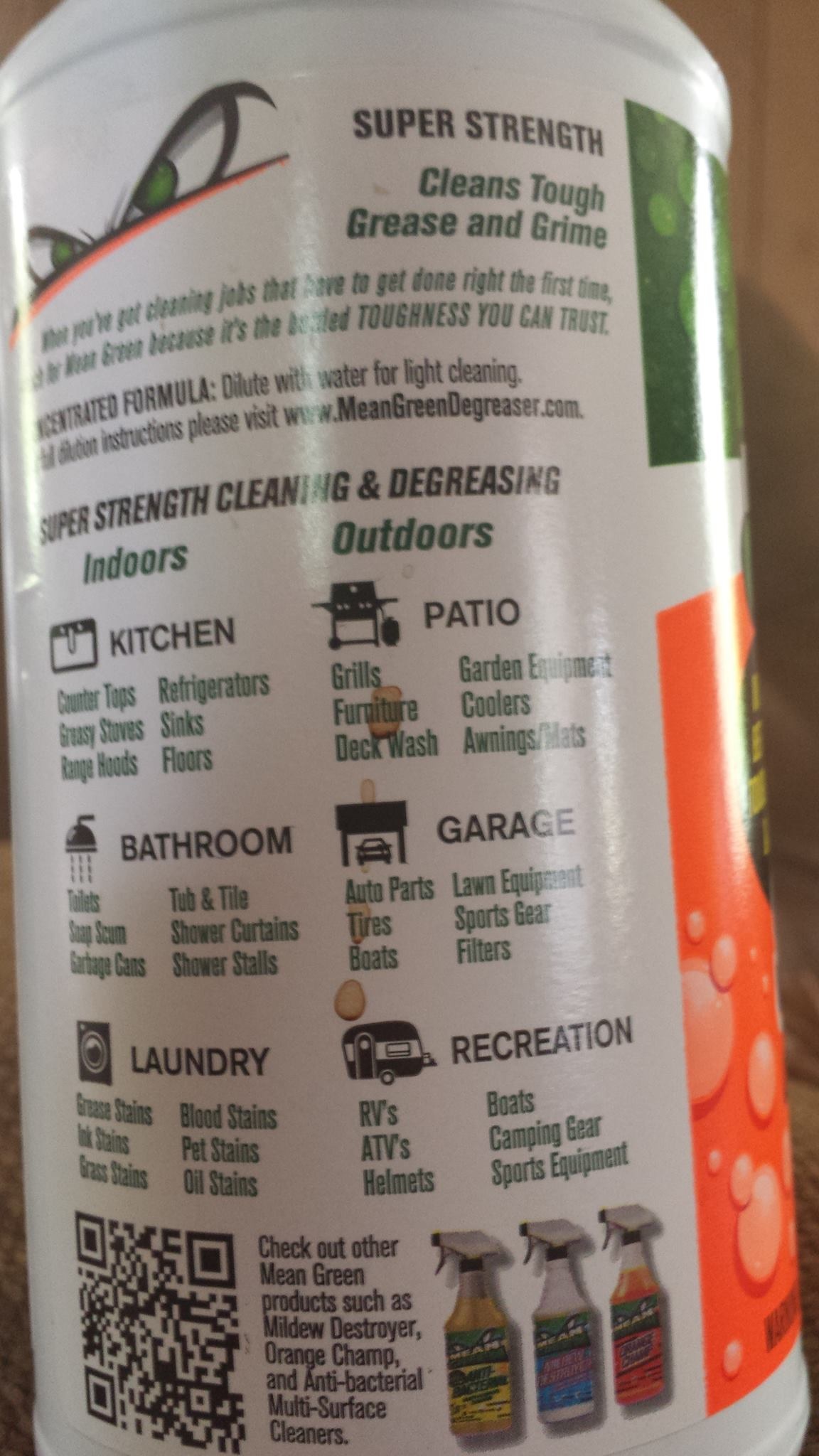
The solutions are many and in discovery .... as are ............................ the problems. ©2015 Steve Beddingfield, being used with permission by Lumigrate. (Photo on right is of what he calls GSB, giant sulfur bacteria surrounding another bacteria. Titled: "Giant Sulphur Bacteria and Its Endosymbiots".
From the longest-standing member of Steve et al.:http://www.menards.com/msds/113535_001.pdf. Later on in the conversation she interjects: Read the MSDS for cautions on eyes, skin, and lung before exposure.
and then a little later from a regular member: MSDS sheets for the products:http://www.meangreendegreaser.com/msds-information
It's apparently in this brand name sold in the US and Canada, other countries have other brand names for the same or similar chemical mixtures, the conversation unfolds to reveal.
From Steve: "A super resistant, giant bacteria is torturing and killing humans worldwide, so I'm looking for ways to kill it." and "Not for internal use; used for home and auto, etc. "mold", which is sulphur bacteria.A wee bit in laundry helps. Skin is a ? mark." He said he'd do a test spot on skin. .... So this is how this evolves and has come to be, this collection of information that I'm providing sometimes the day-of to the pages of Lumigrate for people to find IF they're wanting to use this type of information as a resource.
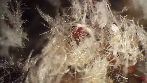
September 14, 2015 Steve Beddingfield added this, after someone reported seeing good results from OdoBan: Physan 20 at Home Depot, in the swimming pool accessories section. High strength Lysol. Odoban is a 2% mix of BAC, Physan 20 is 20%, you can add in your own essential oils but remember to dilute the Physan 20, it's 10 times stronger than Odoban.
Original Eucalyptus Scent
Multi-Purpose Cleaner
Kill Viruses* in 60 Seconds
Kills 99.99% of Germs† in 60 Seconds
Air Freshener • Laundry Refresher
Deodorizer • Disinfectant
Mildewstat • Virucide* • Sanitizer
Active Ingredients
Alkyl (C14 50%, C12 40%, C16 10%)
dimethyl benzyl ammonium chloride......... 2.00%
Inert Ingredients........................................ 98%
Total 100.00%
I have really enjoyed the results and information so far that I've seen from the BioKleen company. Here's the link to their home webpage.... biokleenhome.com/ ... and what you'll find at one of the tabs that gives the history of this innovative business
Natural before “natural” was trendy.

Bi-O-Kleen, born 1989
In the 1980s, with still little awareness about the negative health and environmental impacts of toxin-rich cleaners, one man felt that something wasn’t right. A sales executive for a chemical supply company, at the time, Jim Rimer grew disturbed by the harmful side effects he witnessed among customers and colleagues, from skin rashes to respiratory disease and worse. So when his attempts to implement safety and educational trainings on the use of those products led to his firing, he vowed to find natural, effective alternatives, and Biokleen was born.

Jim Rimer
A Green garage story
Cleaning carpets for seed money, Jim spent his nights studying chemistry and botany to formulate what would become Biokleen’s first leading-edge natural product, All Purpose Cleaner II. (All Purpose Cleaner I just wasn’t good enough for Jim.) In his Portland, OR garage, with only a drum and wooden oar, he designed cleaners powerful enough for the commercial industry he knew. Word caught on about the man who sold truly effective, non-chemical janitorial products, and soon families began requesting smaller bottles for their homes.
25 years of innovation and integrity
 As demand for Biokleen products increased, Jim’s daughter, Cindy, took on the company’s books, soaking in all she could about the nascent naturals industry. With hard work and progressive insight, Cindy moved up the ladder and into an executive, visionary role. She began cultivating retailers—plus a league of brand enthusiasts!—and developing sales and market strategies that helped the business grow. With its move to Vancouver, WA, in 2003, Biokleen expanded its production and renown across the U.S., from farmer’s markets to online stores to individual customers and families. Next Spring, Biokleen will celebrate its twenty-fifth year since Jim Rimer’s first sale, in March 1989.
NOTE, BioKleen uses enzymes, not phosphates, really has the concepts in line with what we're learning here about what to look for. They have an oxygen product. What a way to seguey into OXYGEN CLEANERS!
Oxygen Cleaners - Prepared and DIY Methods
OxiClean was developed by OCI Company Ltd., in South Korea. One of the active ingredients in OxiClean is sodium percarbonate (Na2CO3•H2O2), an adduct of sodium carbonate (Na2CO3) and hydrogen peroxide (H2O2).[3] Related products include OxiClean Free Versatile Stain Remover, OxiClean Laundry Stain Remover, OxiClean MaxForce Spray, OxiClean Power Paks, OxiClean Triple Power Stain Fighter, and OxiClean Baby Stain Soaker.[4]
Here's an example of how the learning goes in a group such as the one Steve Beddingfield has (more about him and the group, below), which has a lot of people who know they have Morgellons symptoms and they're proactive about doing what's needed to research, find solutions, and reverse symptoms. He posted the above information about OxiClean late in September 2015, the last Saturday of the month, and this is what ensued, in a summarized, simplified way I've taken things to put here:
Steve said "baking soda and hydrogen peroxide cant be wrong, thats what OxyClean is, the combo.:
They established that OxyClean used on clothes had helped people with Morgellons, their symptoms are better or gone. One group member said this: "It works for clothes I know that. None of my clothes "crawl" anymore. I used super washing soda, Borax and OxiClean."
They discussed using it internally and on the skin externally (as in baths). Someone said 'another crazy detox bath idea', to which another member swooped in and said "not crazy, its about time people on here learn how to use oxygen. You are suffering needlessly for ignorance of that subject." Then a MASSIVE amount of information came from this member who clearly had been frustrated with people not having been discussing this before. So now that they were 'into it' she was going to get 'on it' teaching 'the class' (the group).
Her words were worthy of transferring for reading here (and I've shuffled things to make it so the more basic stuff is near the top, it gets more advanced lower down when talking about the expert who is named and how these things are used internally but would conflict with the medication used in the protocol that Steve Beddingfield suggests in his protocol (so a reminder here to put in your time to study this information, this is important and not that simple, really, though we have to start somewhere and do that safely and then delve further, we ALL started out not knowing this at some point, remember):
"It is easy to make your own Oxy-cleaner at home. I studied how to do it -- hydrogen peroxide is added at the same time the washing soda: two part formula, easy to do. You just cant store the hydrogen peroxide and the washing soda in one powder like the commercial oxyclean. It is a two part formula with the peroxide containing the over burden of oxygen and the washing soda, which is acidic, acting as an activator. It is similar to the activation that takes place with MMS. Adding the excess oxygen with the acidic activator, and BAM! the oxygen molecules are released into the water, then BAM you have clean clothes or body. It really works, don't pay for the commercial stuff -- it has additives. Using it simple is the best and cheapest way.
... the skin really responds to an oxygen cleaning approach. Technically what makes your skin soft, amazingly, is that the oxygen has burned of and oxidized the dead skin cells that constantly com up on our skilivingn for and cells dying . A dead cell registers to the oxygen as an acidic signature, and it attacks the dead skin cells on your hands, it removes them. No creams can make my skin softer than a bath in calcium hypocholrite, mms, or oxyclean which is oxygen. High five for your skin, get into the healing over all your body. Our private parts benefit for the oxygen cleaning. It is a real good way to clean up.
... well not to get graphicbut baths in this will eventually remove and correct warts!!!
Ive been talking about oxycleaner made at home for months. Calcium hypochlorite, "pool shock", is made of a similar material as hydrogen peroxide and washing soda. It is automatically activated as soon as it hits water. I have been using calcium hypochlorite baths and laundry for months while so many of the people on here suffer from Morgellons lesions. The calcium hypochlorite baths will heal and help, but people on here cant understand oxygen and its uses so they cant see how to use pool shock. Either they want things complicated and expensive -- it's human nature.
You can buy regular baking soda and hydrogen peroxide and mix it . I noticed it's in many of the cleaners now, and toothpastes also.
Washing soda is also called soda ash. It is a different product than baking soda. Seek it out by name: here is some information : The chemical formula for washing soda is Na2CO3, and it is also known as sodium carbonate. It is a salt of carbonic acid, a chemical that produces a wide range of salts collectively known as carbonates. One common source of washing soda is the ashes of plants; for this reason, it is sometimes called soda ash. Sodium carbonate can also be created from sodium chloride, also known as table salt.
You can put MMS tablets into the bath, but first use some tap water to dissolve the MMS ... .... stop screwing around with capsules of this stuff. Go to a pool store and ask for calcium hypochlorite with at least a 72% purity will run you $5!! Then you will have enough in one pound for long time use, make a solution of it. One tablespoon to a quart of water, just allow the solution to settle. Use about 4 tablespoons of this solution into your bathtub -- wonderful.
I dont have the money for fancy stuff; I use the oxygen in many forms. I feel rich for this knowledge, and it has prevented many severe illnesses.
My basic formula it to use one tablespoon of the calcium hypochlorite powder to one quart of water. Shake this and allow it to settle. Use the clear solution, leave the cloudy bits at the bottom. On this amount, for a load of laundry, I use one cup of the clear solution, and I do not use soap either. Important to know that laundry soap is something the calcium hypochlorite solution will fight against. It will get out all soap residues and all dirt and all GSB. Use more if stuff is really dirty, like if poopy. And be sure the clothes you are washing are actually able to stand up to a minor bleaching even though it is a non chlorine bleach effect. I use this on all my linens, all whites, all kitchen, but not my silks or colored clothes.
For the colored clothes, I use a bit of soda ash into the soapy water or otherwise I use only a small amount to clean my colored clothes -- they will fade eventually, but worth it to get out all the soap and poop residues. They come really clean. A one pound bag of pool shock could last a family 6 months of most laundry needs. It is used otherwise in the house in many, many ways I hope to share.
In the bath, about 4 tablespoons of the clear solution made from one tablespoon of the powder, I used more and used less, both are good. I used much more one time it smelled like strong bleach -- I used so much, got into the bath, and immediately a bunch of black seed things left my skin as soon as I got in. Using too much wont hurt you -- experiment with it.
Do not use soap -- this is very important. The action of the oxygen will fight the soap. It will use the available oxygen in the water to eliminate the soap (and the peppermint oil as the question someone asked here was about a peppermint oil soap many have used for generations). Try putting some of calcium hypochlorite solution in a bowl, then add one drop of peppermint oil, within 30 seconds you no longer smell peppermint.
Oxygen trumps terpentine compounds full stop. These are no-soap, no-oil deals -- they get off the film of soap and oil, and of course the biofilm goo that is coming up to the skin all the time. Stay in at least 15 minutes, says Jim Humble
Oxygen is the most alkalizing thing there is because it burns off the objects and substances that register on a pH scale as acidic, like the biofilm, and the acid from the GSB is going to be neutralized by an oxygen exposure."
(Within hours someone had said they used 1 tbsp of hypochlorite in a quart of water and added it to a bath. It felt great... The irritating itchies... redness left It was added to my wash today. And said "Thank you" to this substantial contributor)
More about the expert just referred to, above, Jim Humble, from this featured contributor....
"It seems a new thing -- I was referring to calcium hypochlorite recommended by Jim Humble in his books and articles. On here, most people in following the protocol do not use these products internally.
The stuff which is sticky and rises on our skin is in need of a trimming. The oxygen trims it and dissolves the biofilm. Now, to understand why it is not recommended to take these products internally -- I will tell you why. They could interfere with the actions of the fenbendazole as it builds up in the system. The MMS, MMS2 and stabilized hydrogen peroxide might clean out the fenben while we are trying to build it up. Using mms internally is something people can do with enjoyment, but here we avoid the internal taking of them, please. Using the products externally is actually more helpful to us. These products work well to clean things out internally but fenben needs to build up.
For the sinuses, it is a different matter. I have used the MMS to clean my sinuses and flush them with gas. It gets out the worms very fast and painlessly. Using MMS as a dry gas is the absolute best, most effective way to use that one. I am willing to guide with the gas use, but be discreet and pm me. It is dangerous to the uninitiated, and you can cause lung problems using it indiscreetly. But you know those gum worms people are so upset about? I say gas 'em out. MMS dry gas will not harm tissues applied externally, and in a limited way internally to the sinuses."
NOTE FROM MARDY: If anyone is interested in this, get ahold of me (About Us tab, find me on Facebook, follow information in my signature, below) and I can work with routing you to resources to help facilitate your learning, etc.
(Also, a link provided by another group member about how to make washing soda: http://naturesnurtureblog.com/ttt-turn-baking-soda-into.../
Again, go to the link, above, which is the overall 'here's all the links going out from here to all the topics that have to do with THE Stuff' to find the link about laundry and cleaning and take it to the topic thread at Lumigrate for the full AND updated cleaning/laundry topic about THE Stuff.
The Time is Now
I looked back trying to find what Steve had told me when we met in late summer of 2014, in Facebook message, about his history. That is how I was reminded that at that time he said "next summer is when we'll really find out what this is going to do". I include this here because I want to be showing that even with finding the teacher of the information, I did not 'get to seeing how important it was' until a certain time. Until someone else helping him digest and education in his group presented something that just hit the nerve for me. So I encourage people to know that we're all getting inundated with 'messengers' and information. If I say 'this is extremely important, I think imperative and top most priority, please look', I mean it. So I've said it.
One week ago, I'd 'noticed' three separate places around The Grand Valley of Western Colorado where I saw what appeared to be white cotton candy, or haunted house cobwebs, or spider webs all over a bunch of shrubbery. All of the sudden, it just seemed like either I was knowing what to see and now 'noticing', or there was more to see and notice. Yes there was a spider nearby two of the three and I could imagine how the third had a spider that had wandered away and didn't die there. One was small and stuck to a little 'webbing' and when it fell apart, I counted 11 legs and maybe more had crumbled. Bizarre, maybe more --- or less -- after having studied the information Steve's taught thousands now.
I NOW know that what I thought in the past were just spider webs in the grass, appearing overnight and baffling me several summers ago as to why we had so many spiders all of the sudden was not what I thought it was. The way the cloning and DNA transfer happens with THE Stuff, giant sulphur bacteria, allows for explosive numbers to occur. By what we thought were algae blooms, which actually have GSB (THE Stuff), there will be massive numbers of a progressive neurological / neurdegenerative disorder OR another. Not some of this and that and the other.
This is because it's getting the 'stuff' from an organism that causes a disorder and replicating it. So people and everything in the area has gotten exposed to it. And a 'cluster' will have sudden symptoms onset in the time after, the wheelchair vendors then are baffled as to why it's ALS here and MS there. Until they learn this theory and then 'that makes sense' is what they say.
I'd lived since the spring of 2006 at the place where I saw the massive 'spider webs' in the grass one day. "Quotes" because they're not spiders, they're spiders that have had The Stuff mess with them and THEN replicate. I wish I remembered what summer it was, maybe 2012, when I saw them.
Soon I noticed chemtrailing start (a little in 2011 but it was massively, daily started June 1, 2013), and I knew that was imperative to learn and tell others about as well -- it had to affect health, obviously. It turns out, according to Steve Beddingfield, the chems in the chemtrails are aimed to help combat THE Stuff. THE Stuff is giant sulphur bacteria, or GSB. Yes, they know the stuff is falling to Earth and getting in the trees and waters, and into people, but it's hopefully going to help the problem and overall the lesser of the 'evils'.
You'll likely want an introduction to what THE Stuff is about, so I'll provide it here. I'm sorry, it's mind bending stuff and so it's not going to be that easy or fast but I assure you I have worked to get a version that is as easy and fast as well as interesting and 'digestable'. If all I did was say 'problem, example, solution' you'd feel overwhelmed.
I will also present the categories of areas of our environment and 'lives' that this affects; we'll have examples and, if possible, photos sometimes. But this will be solutions-focused and I will be turning to my contacts, many of whom are Facebook friends.
YARD/ OUTDOOR SITUATIONS
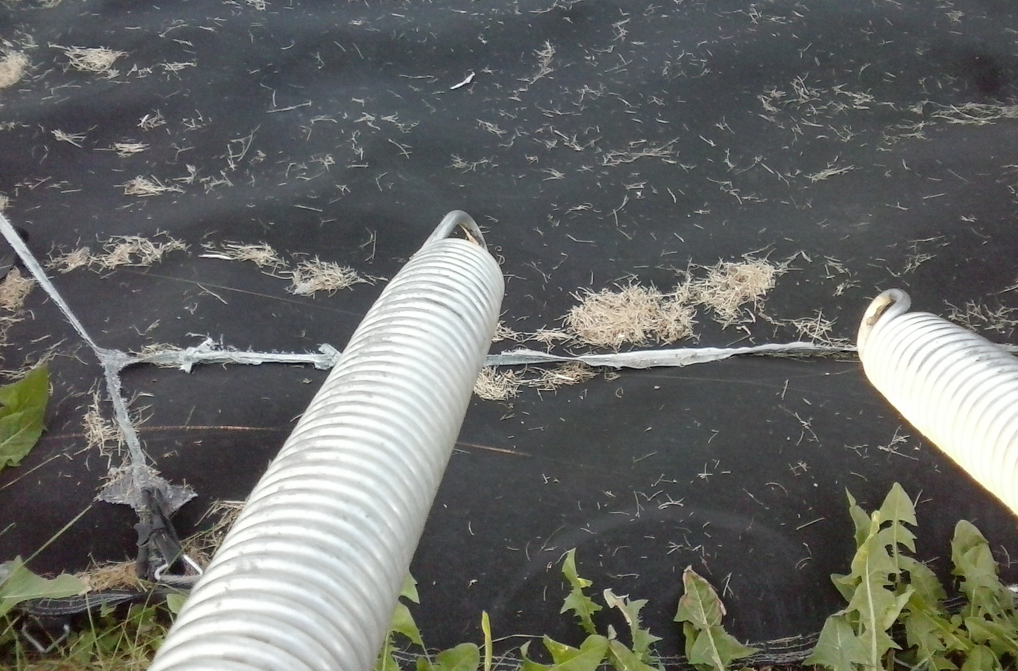
Glints of light on the fine strands, two of them, visible off the left spring and extending to spring on right (visible) and left, off camera. The days surrounding when this picture was taken, there were strands also seen spanning across this out-of-commission trampoline frame, one longer than five feet, the other about eight.
Clothing -- Indoors or Outdoors
© 2015 Steve Beddingfield "Tubules" -- This was on the floor of my buddy's hot rod garage. It was on some 90 weight gear oil which is rich in sulfur, which makes it smell like what is referred to as 'sour crude'. The Gulf of Mexico always has sour crude.
Tips about doing laundry with products and processes that are more beneficial at remediating THE Stuff can be found at this topic previously created at Lumigrate. I've worked in-home with people applying this information to how they do their laundry and helping reinforce the information.
NOTE: If you have a communal situation with other people using the equipment, you might look for someone you can help 'do something' in return for using their laundry equipment. If you have enough TIME to hang out and do their laundry, with your products you provide, or something else you can do in their home when your laundry is processing. Problem-solving the problems in situations is what occupational therapists were specialized in and I have that as part of my background (graduated 1996 at age 36, a 'second career' and this is now third).
Prevention
Keeping the stuff off of your skin and out of your hair becomes a priority to suggest people consider doing. Realistically, thinking it through, this information is going to change hair styles. We should consider keeping our hair 'minimized', and the same with body hair. However, the trauma of shaving might exacerbate and provide an entry point. The bacteria, with its
Clothing: 2100 count polyester and minimal uncovered skin, such as the face, similar to speed skaters.
Mask for certain
Cover the head
Wear long sleeves
Wear long pants
Boots -- limited exposure of skin.
Place clothes immediately (ASAP) in water with OxiClean and then launder according to the information provided at Lumigrate .....
The dryer lint will be toxic as well, so it should be handled with that in mind and disposed of, ideally, by being burned and the ashes are then something to figure out what to do with.
I realized I was thinking how my dad had coveralls he'd put on to go outside and work around the property. I was in the habit of wearing jeans or shorts and whatever shirt, but in the summer it would be a tank top. I've been 'wary' of things where I am currently (CW Colorado is appearing not 'as affected' but I did see my first 'feather' looking tubules in July. It looked like a very small piece of down and was tethered to each arm of an outdoor chair by long, thin strands that you'd think was created by a spider ... but how would it have done THAT? The chair had new cushions just 'out of the box' from a big box store a few days earlier -- did it have anything to do with that? Who knows.
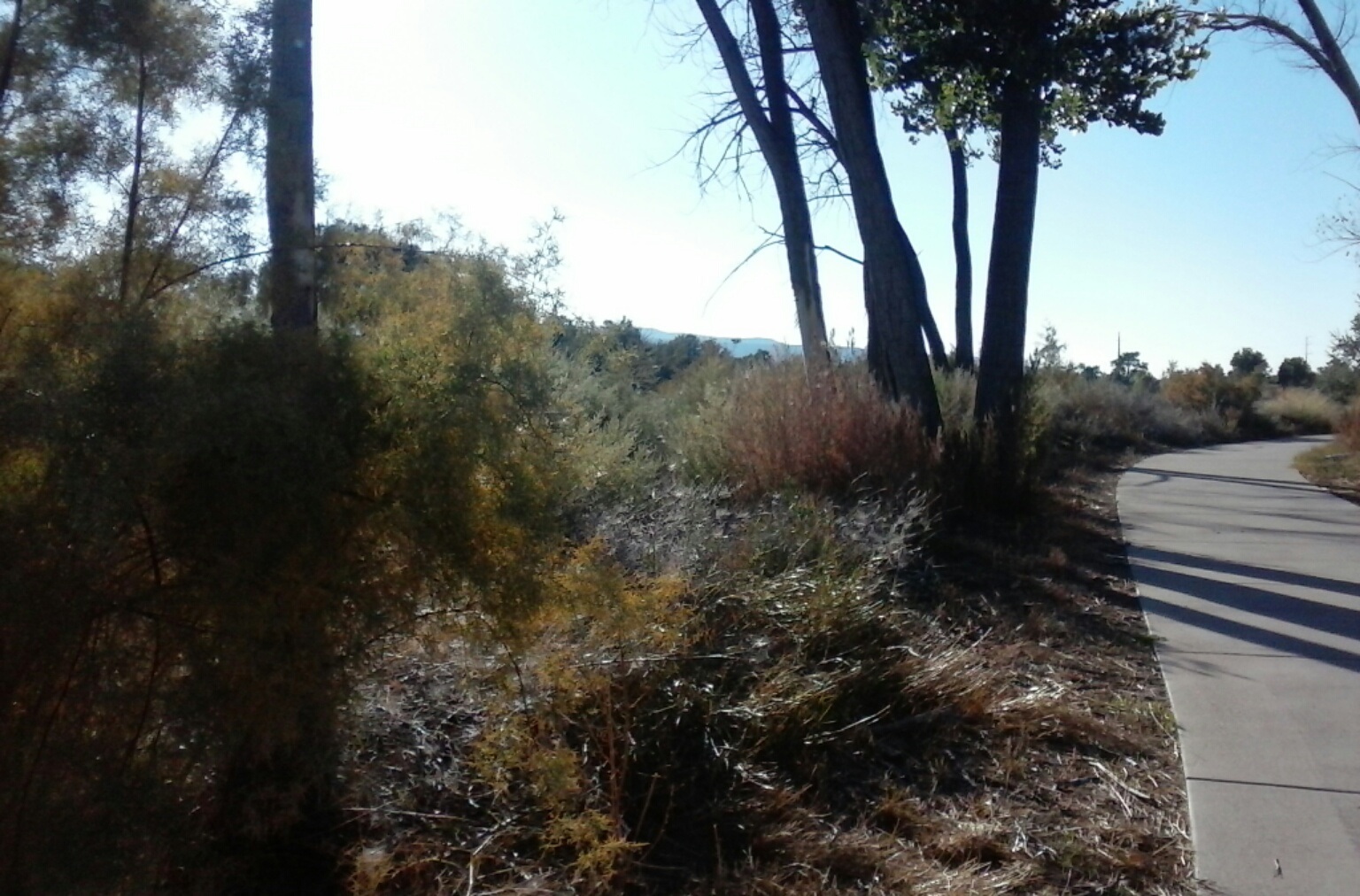 Sept 7, 2015, Colorado Riverfront Trail showing stressors and dead foliage with much white-appearance similar to in junipers, photographed previous month. Showing it more 'close up' and sun shielded, below:
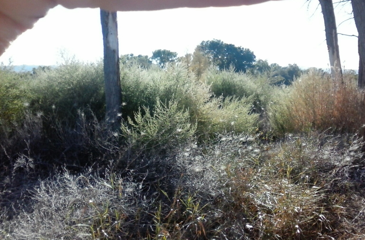
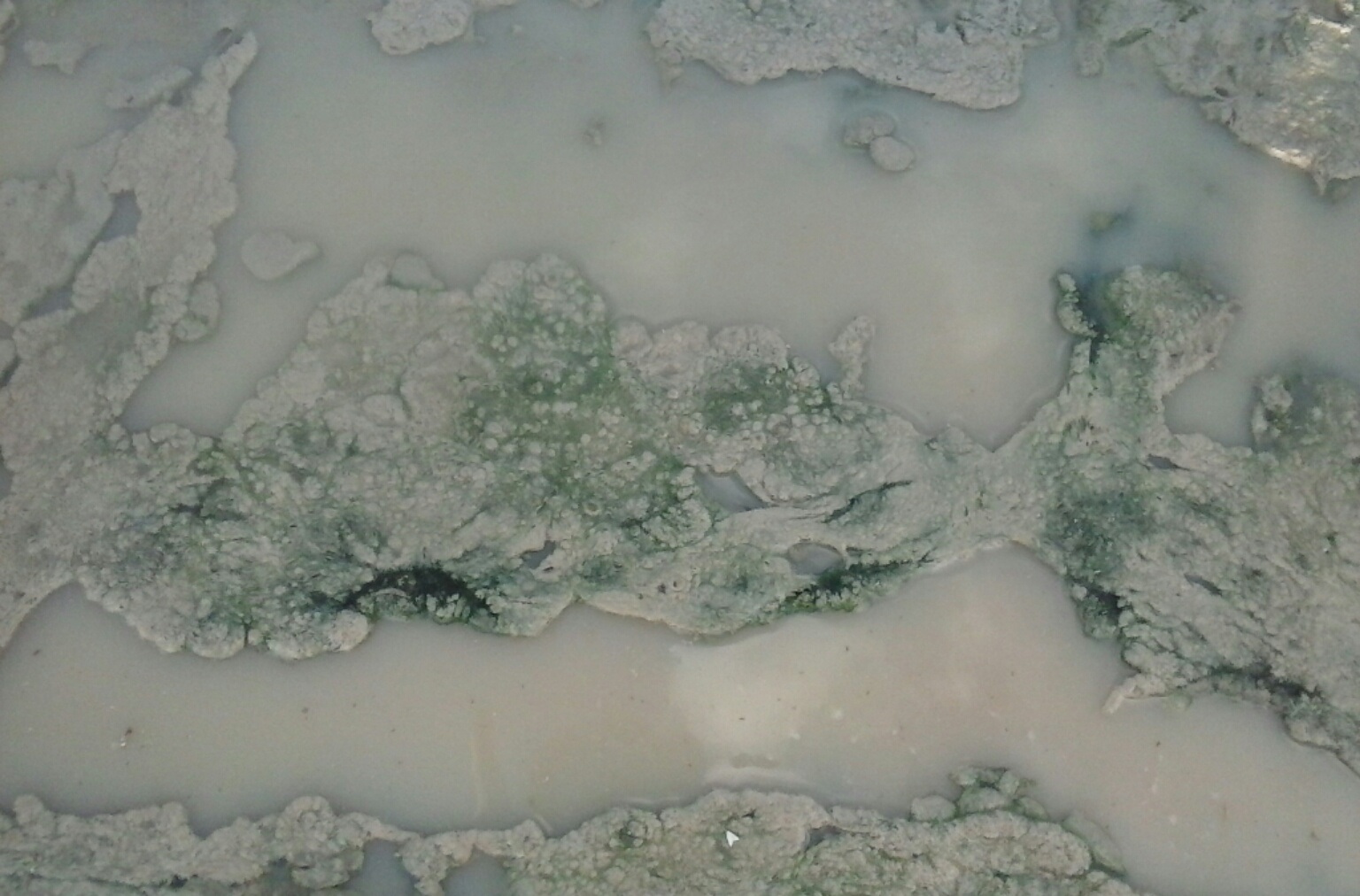 During the same week, in the desert, within 1-2 days after rain fell for the first time in quite a while, this was in a rut that filled in with water. The animals walk through this, and it dries, becomes airborn and then settles elsewhere. In the same timeframe, I witnessed the long, thin hair-like strands of what Steve teaches is GSB on plants, buildings, and found I was getting it on me as well.
The dog started being very bothered with things that both made it scratch and chew, even suddenly dropping to the ground to start tackling something it was experiencing sensations from. Treatment to the rescue, thankfully knowing but reflecting on my dog from the 1970s who lived to be 16, had a tumor much of her adult life under the skin ... just one .... who'd responded well to something sulfur topically applied. But the misery these animals are going through -- human and otherwise. .... I hope by showing things I observe personally, it will help YOUsers of Lumigrate connect dots.
In walking in the same area later, I noticed that once the water was gone and this area of the rut dry, there was no trace of green that I could see, but there was a greyish color that would not alarm me in terms of what it was.
COMPOST, WORM FARM, SOIL, VEGETABLE GROWING
From "The Worm Farm Wizard of Oz" said this with his posting of about 15 photographs (I've only taken one to show as this is going to be a very long thread I am sure.) "I put a bunch of fallen leaves; Some dirt and torn up paper into my worm farm weeks ago.... after tipping out most of the worms into a few over-filled compost heaps. Tonight pulling back the paper to pour some rinsed out soup pot into the worm farm, I noticed and photographed these at 1x and 4x magnification. I'll post these under a similar comment in the group. Thoughts on this being GSB / cyano-bacteria colonising the mould process in the breakdown of the leaves?"
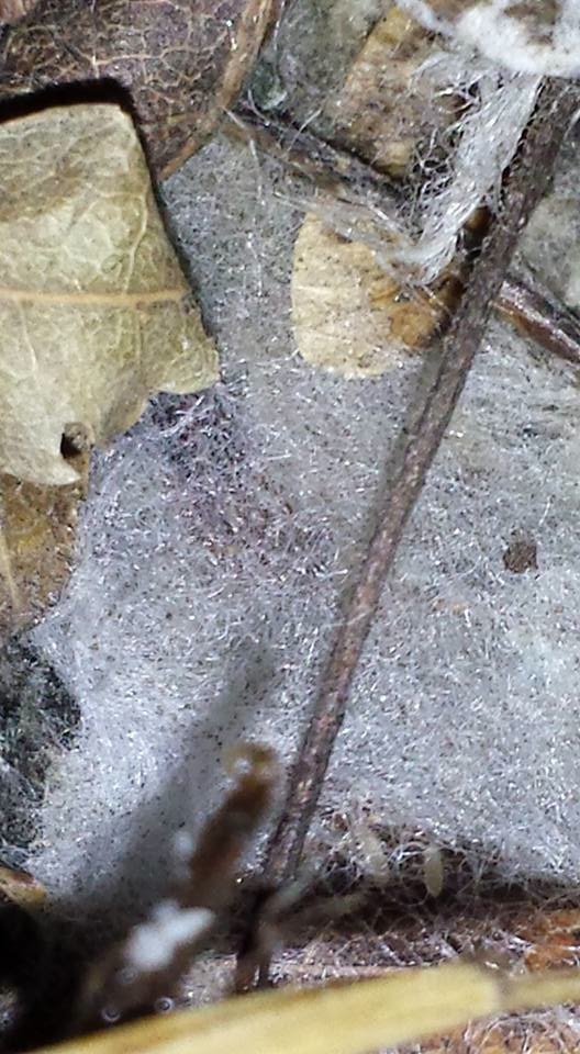
RETHINKING OUR PRACTICES of how we decompose our organic matter growing in our gardens, yards, and what we compost or otherwise dispose leftover biomass. Everything will need to be treated, in ways yet to be determined, and put back into the soil and onto the Earth. In the mean time, doing things differently -- keeping things dryer not moist, so as to reduce the growth EVERYWHERE inside, outside, EVERYWHERE is the key concept. In the past, without THE Stuff as a factor, we'd toss in the garden or 'field' the fruits and vegetables (which hold a lot of moisture) into the soil or into the compost. Now, as a first step, drying those as much as possible and perhaps incinerating them, then treating the ashes.
BioChar is a concept which Steve Beddingfield is intrigued with and believes will offer the solutions. Essentially using a process similar to making charcoal with the biomass, then adding enzymes and potentially polypores is an idea that Steve enterains when discussing this aspect on August 22, 2015. Come back for updates, as he's perhaps going to do more thinking and researching and talking with others and bring new ideas to the table, that is his "M.O."
More from the Australian soil-focusing guy, September 6, 2015.
" I planted up some runners of water cress last week....basically garden soil composted with food scraps and compost words and old wood fire charcol mix as a cheap potting mix and old styrofoam boxes."
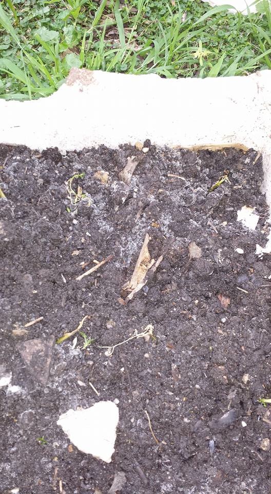
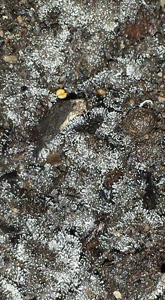
Left = 1x magnification, Right = 5cm away and 4x magnification. Greyish whiteish fur growing faster than my water cress...havent seen it do this before. New or newly-noticed, I cant say. But wonder if i should toss it all?
Steve Beddingfield: Deadly! Toss it! The soil requires bio remediation with fungi. Polypore said (sic.) can be grown on logs, I like digging a trench, placing the logs half submerged lengthwise, inoculate the logs with cultures of fungi that are specific to the wood. The wood chips can be used for growth media, then turned back into the soil.
The Aussie/ Oz-ie guy then said: "This would suggest all the commercially grown foods I've composted have it and it is there waiting for the ideal conditions. It looks like we have some serious remediation to do around here. .... Then he shares they're moving in two weeks (because, I might add, he becoming aware of this STUFF recently, and I want to toot my horn a bit that I'd encouraged him to get active and focus on what Steve Beddingfield was teaching in his Facebook group over the winter of 2015 for us in the N hemisphere (it was summer for him down there 'down under").
Naturally, much appreciation for Steve's having created the place for learning and then for me to harvest information to take out there to YOUsers on Lumigrate and in my work educating about what's causing health issues and what we can be doing about them! And to all who are working to learn, supporting Lumigrate and my work in whatever ways you're doing so. Thank you, and remember, we're all in this together!
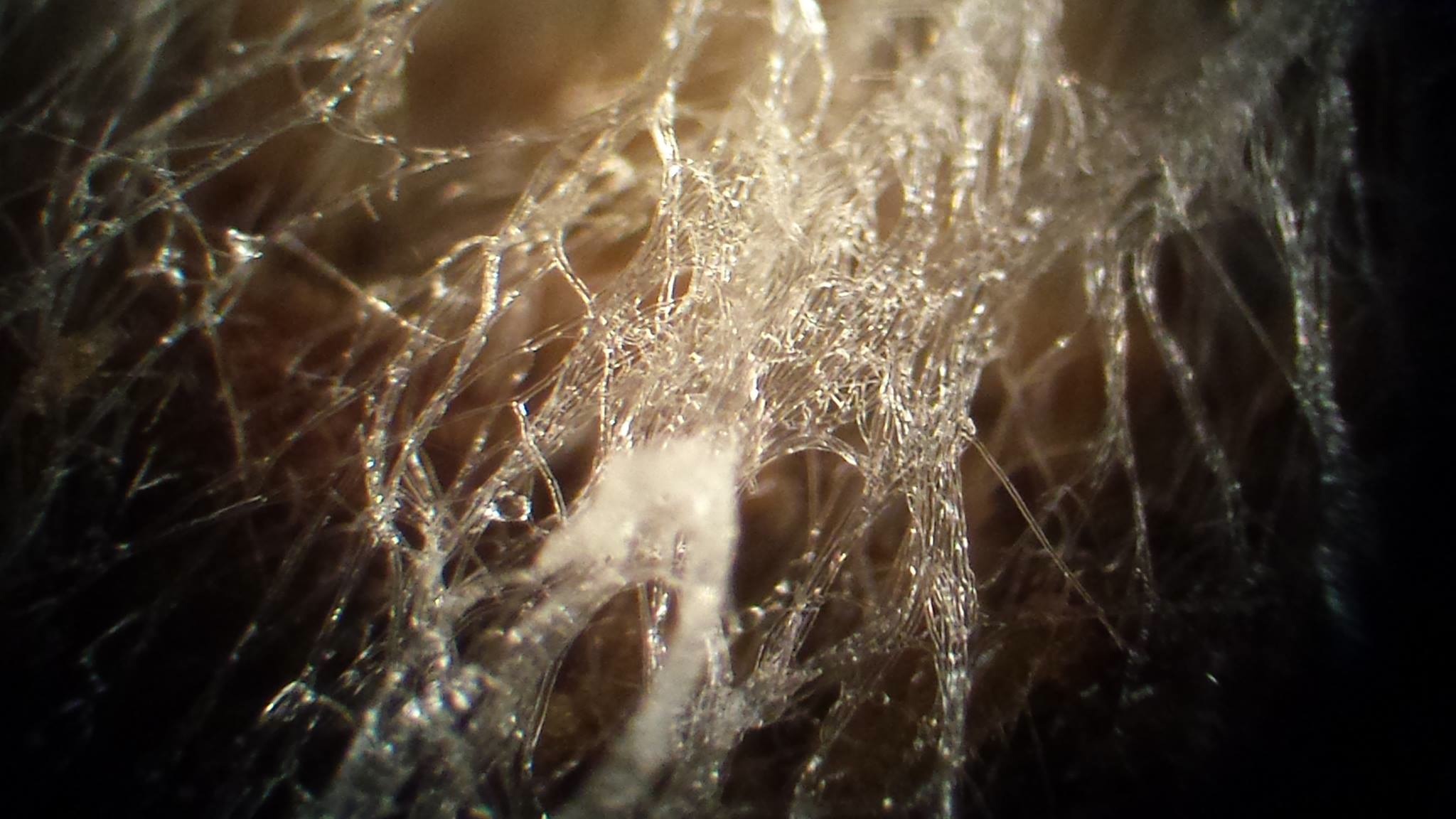
Sulfur Bacteria, hollow filaments wet with hydrosulfuric acid. 60X mag. Found lying on the grass. Dangerous, pathogenic. ©2015 Steve Beddingfield.
BUSHES, LANDSCAPING
© 2015 Mardy Ross "THE Stuff in Landscaping, Grand Junction, Colorado. August 2015"
There was a spider / "spider" in the closest one to me and the camera, but I didn't see any in the other "webs", or "masses of weblooking stuff". IDEAS TO CONSIDER, via Steve Beddingfield, for remediating this:
See above relative to biomass in general, which presents a new problem in terms of the bacteria remediation. Clearly, removing the affected plant material (and getting it away from the buildings) is going to be preventive maintenance. MINIMIZING what you have for this to be getting onto and getting growing into that becomes airborn, gets to the soil, etc. is going to be a fundamental concept.
Trees
What is affecting trees is now more complex than we typically have previously known. This is a paradigm shifter. You'll see at the topic I created at the blog tab which links to every topic that has THE Stuff as major content for following to for thorough learning that I cover information about trees there. Here's the link (provided also, above): www.lumigrate.com/blog/what-i-want-every-mother-and-others-know-then-go-there-cause-all-problems-presented-emerging-re
For this 'introductory' topic, I'm trying to provide fewer things which help people make the massive paradigm shift as easily and fast as possible. Not that it's easy, this is really difficult to wrap your head around, as I say repeatedly. But here, I wanted to provide this photo that Steve Beddingfield provided on September 7, 2015, Labor Day. I worked Labor Day too.
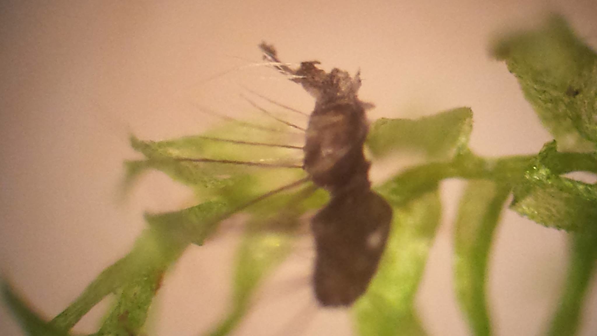
©2015 Steve Beddingfield Found in a N.C. tree on "algae"-appearing material. "Sea horse perhaps, from The Deep"
In The Home
Appearing as 'dust bunnies' or 'cob webs' or 'silk threads'
I made this photo large and also sent it with full pixels since these are so thin and difficult to see. Without the right light, as shown in this picture, these thin strands were 'not noticed'. Refer to the photo, above, with the springs, these were taken on the same property. What helped me to put the pieces together after learning of THE Stuff, was experiencing seeing long thing strands airborn outside, floating along, wrapping around the tools I was washing windows with in 2013 and then this year I remember one occasion opening the door and saying 'hi' to someone and then both forearms felt like something very thin and hairlike was on it and sure enough, wrapped all around my arms .... not spider webs 'blowin' in the wind', apparently GSB.
The Kitchen
Produce Preparation  © 2015 Mardy Ross
First and foremost, become an educated consumer and look for providers who are as well, about THE Stuff. Know which vegetables and fruits typically have more or less of THE Stuff and make your choices of what to consume with that in mind.
The above vegetables were picked a week ago by The Produce Peddler, and have been taken to the farmer's market and then out on the counter for a day and then into the fridge BEFORE doing any post-treatmentby me.
Clearly, it's 'ideal' to get your produce or produce your own, using people who have learned about THE Stuff, know what to look for, and are working to be on the cutting edge of wellness of the soil in regards to THE Stuff. Ask them to cut something open, you can sometimes see THE Stuff, as shown in other photos provided here, with the pickle. Steve Beddingfield stated that a person can take a 'locking' type of bag and put a vegetable in it with as much air as you can capture for it to have something to get growing with, leave it in a safe place for about a week and see what grows. You can put things in a cup with water that's pure, if you are fortunate enough to have figured out how to have pure water, inside a bag with air before closing it up, as well (for things that don't have as much wetness). He suggests doing this with the brands of things you're purchasing, and if you're growing produce or are otherwise obtaining produce, you'd benefit from this laboratory experiment / exercise.
I'm wanting to help those growing food whether for their own use or to 'peddle' / sell learn about THE Stuff, particularly if it's food I'm going to be consuming and having the effects -- positive and negative -- from whatever's on and in it. And I'm hoping those who USE food, learn and do what is needed today to consume food at it's safest for our long-term, overall well-being. "Lighting the path to health and well-being" is Lumigrate's motto, as seen on our logo.  Ideas here.
How I Went About Making Breakfast Recently, Using My Current Knowledge and Tools
I got these vegies out to make breakfast with (sautee in quality butter, use a little water and a lid with eggs to 'baste' them, it's nutritious and delicious, and utilizes these 'here now in abundance of the harvest' fresh vegetables, which gives you a lot of different things with it different from the carbohydrate sources people have typically had for breakfast. The rest were pre-pared for snacks for the day.
Learning about ozone, as I did from PosterGal and other women when she was a member of Steve Beddingfield's group on Facebook at a time they were reveling in the amazing presumed or perceived results they were experiencing from ozonating their family's produce (and making their water "pure", or really likely best to say "purer" with it as well), I have generally stopped using "The Clorox Method" that I'd previously put on Lumigrate, which I'd learned of from a well-known nutrition expert's book (The Fat Flush Plan just sounded gimmicky to me, I'd only read it after an MD expert on Lumigrate suggested it in 2012, and I found a copy at the lovely used book store in downtown Grand Junction, Colorado where farmer's markets are 'the thing we go and do' increasingly).
When people are working on reversing symptoms, much has to do with cleansing out the organs of elimination from the body, and naturally you'll want to be utilizing food that does not add to your 'load'; load theory and full barrel syndrome have been conceptual models used since the start of Lumigrate. You'll find them BOTH shown on the link in the box, below taking you to the topic about ND Christopher Lepisto's information about what he suggests for people who've lost their wellness and were identifying with the form of illness labeled by those who do so as 'fibromyalgia'.
You'll find that the information Steve and his followers and helpers have come up with shifts our thinking to that we all have one root cause, one 'thing we all have', and it just takes different symptoms with each organism. In people that can be more physical symptoms, or more mental, behavioral, and even affect spiritual abilities and wellness. It's not an easy paradigm shift due to how long most of us have steeped in the illness models and learned from allopathic science and medicine the "A to Z" disorders we need a label gun to keep up with marking what all we present with symptoms of anymore (some of us). See the information, below, under And This is How It Happens. Wanted to just 'prime the pumps here'
I have a few topics to refer you to for more about that if interested in water, the Clorox method, and the basics of how a good naturopath would educate people who have lost their wellness to reverse symptoms, back in 2011 that is -- I'm sure he'd say things a little differently today but I feel it was one of the best overviews I've heard for those who are with significant symptoms. And I have suggested this link to those in Steve's group who are struggling with their symptoms and detoxing as they 'get this stuff out of them'. Incredible the continuum of what's inside of people, depending upon all the things that 'load' and 'unload' a person, whether envisioning a barrel or an elevator as we offer at Lumigrate as visual aids/ models.
The Clorox method topic at Lumigrate (which had only 4,000 reads in the two years it has been on Lumigrate so far) ... www.lumigrate.com/forum/cleaning-food-vegetables-fruits-eggs-even-some-meats-simple-chemistry-rescue . You'll see I say this there: "
I was surprised to initially learn about this on page 134-135 of The Fat Flush Plan, by Ann Louise Gittleman, M.S., C.N.S., in the Chapter "The Master Fat Flush Shopping List", that way back in 1988 when the author wrote/published her first book, Beyond Pritikin, that the common household product, Clorox bleach, could be used as a food-cleanser."
My ND from 2006 and into the years forming Lumigrate when we were both part of a clinic 'under one roof', interviewed by someone on the East Coast and I eventually transcribed or overviewed what was said, included graphics and other links to refer people to more detail about some of the subjects he introduced in this excellent, easy-to-listen-to podcast: www.lumigrate.com/forum/my-interview-naturopathic-medicine-cfs-and-fms-cinda-crawfords-website
Water is the focus of more than one topic at Lumigrate, but this is the one I hope EVERYONE finds: www.lumigrate.com/forum/water-key-environmental-wellness-or-illness-more-important-today-ever-many-aspects-consider -- please see Comment 1, from May 2015 (from me), about Joseph Bender's guest appearances on OneRadioNetwork -- I also overviewed / typed out what was (mostly) said for ease of use and referral.
I find the ozone easier than the Clorox method, though it does take electricity and there's only one gadget at this time since I've only purchased one. So I'd learn about other methods, try them, and have the things on hand. Some use food grad hydrogen peroxide, for instance. You'll find information about that in the 'master's level', long, complete document about Steve's group, research, protocol.
Rather than just taking vegetables you purchase and cooking with them, as I've seen people do (not even washing them with tap water, and any kind of product to get 'things' off that are on the surface), if you're wanting to reduce your intake of THE Stuff (and chemical residues, and etc.), process the vegetables.
This is what I do: Put the vegetables in a stainless steel bowl, you can also use glass:
fill with water that's without minerals (I use RO -- reverse osmosis), and then ozonate per the instructions in the manual that came with the A2Z ozonator I purchased in May from the company. I have been known to cheapen the energy and money expense of water by using filtered tap water for ozonating vegetables but that's based on the relative quality of the water where I am.
(Most in Steve's group who purchased an ozone generator had gotten this $70 (new) model from Amazon, which has free shipping; I was bundling mine with an order from someone learning through my tutoring in-home about this information. I like talking to the companies that make things, as I've said here about food growing companies. Ozonation has quite a lot to learn about it, such as if you ozonate minerals they will be oxidized. So learn about water, it's been covered and I'll not go into those details in this '101 level' topic. 
© 2015 Mardy Ross A2Z Ozone Generator, Produce in Stainless Steel Bowl
In the case of speaking with the founder of The Produce Peddler, who impressively studies a great deal, I learned there's a farmer in the San Luis Valley of Colorado who is ozonating water before applying it to his produce/ crop! Now THAT's my kind of Produce Peddler, THE Produce Peddler of Mack, Colorado was a 'stand out' from the crowd of vendors I've spoken with so far this year in this area. Here's a link to their website, and they mentioned they DIY and are loving every aspect of running their business -- marketing included. www.theproducepeddler.com/ ... Why do they farm? This is what they say:
Why we farm
Our mission is to grow, market and sell quality produce to the people of the Grand Valley in a safe and responsible way. We respect the land & environment and continue to educate ourselves with the latest advancements in sustainable agriculture. The Produce Peddler is passionate about increasing the availability of affordable, fresh, local food to our community.
I'd add QUALITY to that last sentence based on what I found with the produce, clearly, and from walking with Zay and Leah Lopez. This is just ONE example of how to check into and talk to a 'produce peddler', I encourage YOU to do the same or what you feel is right for you.
I did the same thing with meat, as I am not a vegetarian. I will give a shout out to Jubilee Family Farm from Olathe, Colorado and look forward to continuing our conversation about this now that I've had more time to talk with Steve Beddingfield about what these food producers could be looking for in order to know about THE Stuff and provide the safest, best quality from that perspective, given this 'new' to us information.
And so on ... dairy, cheese, etc. ..
Drains - Sinks, tubs, showers
Clearing sink, tub, and shower drains which, in the opinion of one of my 'mentors' on this kind of thing, are "likely the number one source of contamination." She tries to put some Borax, OdoBan, baking soda, Oxy Clean, vinegar, Pine Sol (or similar brand), Rid X, MMS/CD, hydrogen peroxide or Kleen Green in all the drains before bed and let that sit overnight. "Some of us have MCS (multiple chemical sensitivity), so will need to find what is the least toxic, best tolerated, and most effective. I find this measure makes a huge, huge difference in how the house "feels" as far as livability goes."
Someone I got 'into' this information in the spring of 2015 had this great input to add:
"A tool I have been using is www.flylady.com - a wonderful program that has been set up to work in "zones", prioritize and customize tasks, working in 15 minute spurts (she's big on setting a timer). She also emphasizes taking rests for 15 minutes and drinking a glass of water during the break. Also a "weekly home blessing", she calls it, kind of doing a once over of the general tasks needed across the home before you work in your zone for the week.
Printable checklists, lots of tools available on this website, and I believe a great tool for managing the STUFF. (And to add, great for someone like me who has endured neurological and psychological effects from infection - good for routine, discipline, ADHD symptoms...) For example, a great customization for your zone would be to ozonate whichever room you are focusing on that week (leaving it to one room at a time, not the whole house - so if you are housebound, you don't have to worry about having to leave when ozonating (very important) and end up not ozonating because of that important safety factor of not being present when ozonating).
Ultimately, once you make it through all the zones once, you'll find that the routine becomes easier and you'll see less and less of "the stuff" in your home as you are cleaning. There is also an android app for this program that incorporates the checklists, and is very easy to use and customize. It is called Flyhelper. (I'm not sure if there is an iOS version or not.)
The website will lead you to 28 days of what is called "baby steps" - this is for the people who need a little push in developing new habits, and the concept of it taking 28 days to make something a habit. The shining the sink thing, she's onto that for aesthetics, but if you think about it, really is important for us to do daily when addressing the health aspects of cleaning and mitigating all this stuff.
I hope this helps some with cleaning. I thought a couple months ago that the cleaning would ultimately kill me or the stuff would, but now I feel better about it. I'm currently mostly using the Odo Ban and following these procedures.
A woman in Europe provided this link, thankfully, as it has tons of interesting information focused upon flies being a component in chronic health problems, much overlooked: http://www.morgellonsuk.org.uk/micromyiasis.htm -- The part I pulled out to put focus upon here:
"Morgellons sufferers are very aware that their clothes, fabrics and homes are affected.
Sufferers often report finding fine hanging silk threads and webs spanning furniture and wall surfaces. (Mardy's note interjecting, I'd say silk-like and web-like instead....) Common reports are regarding fine webs that have an irregular construction (ie. not a cob web). Interestingly, the small cob web spider produces irregular shaped webs to increase their likeihood of catching tiny flies."
I did not know that about cob web spiders, I thought any kind of web that was in a place you'd want there to not be webs would be called a cob web. Learning all the time, I am! I hope YOU are too!
The Heating, Cooling, Air Circulation of Homes and Automobiles - Basics
One has to consider the materials that you're cleaning, it's important to remember so I'm going to lead off with that. Ductwork for forced heated and refrigerated air, for instance. The filters, what material is that made of. Or if it's evaporative cooling, what are the pads made of?
There's a lot to consider. Best if a person learns the basics, puts on their own thinking cap and gets some ideas. And gets a microscope, and the type Steve Beddingfield has suggested to people is listed in the topic that has extensive information about his teachings from his Facebook group. About $100 or so, better ones when you get more into it or are commercially interested go up from there in price, attach to the computer and you can take photos and videos, and etc.
For others, they might consider contacting Steve, making a donation to his research efforts and sending things to him for having him take pictures of, try various cleaners he has on hand on, or certainly sending along whatever you're wanting to have trialed might be an option too. if it's not the basics he has tried.
Purple Power cleaner in the automotive section of WalMart or other automotive places or elsewhere is one he often refers to. TSP, is one he mentioned to me about cleaning the vents for air conditioning and the outside unit. Lime that used to be added to whitewashing, and can be put on the ground, is another. Again, there's more information about this on the subsequent topics you can go to from here for more details.
The bacteria's biological processes make them inherently sticky, so you're wanting to break that down to make it easier to remediate. You're wanting to protect yourself from getting it on and in your body. "It can kill you" he has said to me. He found a bail of hay on the roadside, per one of his many stories, and opened it up and got blasted by toxins which made him swell up and look like the Lion in The Wizard of Oz in a photo he has on his Facebook or in his group.
The woman who I mentioned and quoted, above, who uses the software to keep on track with cleaning regimen, is clearly a very hard working learner and do-er, who has just taken off 'into it' and improving her health, or at least addressing the new exposures better and staving things off for self and family in her home, where she can control things a bit. Propelled by her own health relapse earlier in 2015, then having death hit close to 'home and heart' visited the graveyard where someone she knows was recently buried. Late 30s, breathing issues but nothing in the system but the correct amounts of prescribed medications.
So she "noticed" the relatively young ages of those buried there recently, she investigated what she could find online and there were an extremely high number of unexplained causes and overdoses. Overdosing perhaps requires some rethinking because there are a variety of ways this can occur: it essentially is a matter of the mix of chemicals the body has going on overwhelming, so that's a 'bar' that changes based on the mix of what a person has taken or encounters unwittingly, and has going on in them overall.
There are a lot of opportunities for people to be doing the work that others cannot or will not do; those who have had less problems with the exposures to date are going to have a different outcome from being exposed than those who have had more. So just be thinking about that as you go about addressing things. For your wellness, you want the stuff managed, handled, remediated, addressed -- but how's that going to be done?
And what I've seen, the people who have had the exposures from their environments in my area of CW Colorado being more toxic seem to have their wellness levels taking their abilities for processing, caring, thinking about this 'extra stuff' to a level of concerning delay and I almost imagine that 'it's a trick by the bugs' to make people want to be distracted by what entertains them. Then if they do talk with someone about doing the work they have to know the information well enough about THE Stuff to convey it to the potential workers. Or they can certainly provide this information if they're aware of it and if the worker will look at it. That has proven to me to be a frustrating challenge. The 'overwhelm' that professionals have in various industries has prevented much study IF they're of the mindset to be open and interested in it, which not all are. So it goes. Again, there's a reason why I put the photo image of the Serenity Prayer on this topic! Even keeping Steve Beddingfield on topic on the phone to talk about one situation entailing an air conditioning situation for someone I'm helping with advise about applying this Stuff information to -- he wanted to be talking about what could be developed, not what we need to know right now for this one situation, which can then be utilized here for others to learn.
THE Stuff: 101 - Introduction, History, Basics
I know YOU are busy. So am I. I believe I have 'leading edge information' here, and it's something that's been affecting our wellness, literally 'forever', but has been increasingly caused problems in modern times. The reason I am SO busy as I prepare this, in early/mid August 2015, is because with the summer's heat having gotten things 'really growing and going', and the cooler temperatures starting which means the water in bodies of water turn, which is when the algae blooms, we're potentially about to see environmental illness in animals, people, plants, insects, etc. go 'off the chain'. I think I'm seeing it happening. I quote Penny Lane in Almost Famous: "It's happening....."
"Everyone" is talking about the numerous things around them 'standing out' -- 'everyone' is talking about bone cancer it seems' as Steve Beddingfield said recently, or 'everyone here knows someone with a mysterious death lately' as someone in his group just said today. The infamous PosterGal had said on her wall today as well that she's seen it 100 times, children needing intestinal surgeries -- their intestines are just dead, black, bad. This was in response to a woman FB friend of PosterGal's who said she had to have a kidney transplant before she realized about functional medicine's looking at underlying causes and so now she cannot take many of the things used to fix the intestinal biosphere problems because of their impacts on the immune system and having the rejection issue with the transplanted organ/ kidney. People who don't have health issues nor focus on that on Facebook have told me just this summer they've noticed many middle aged people very ill or dying or dead. "It's happening", as I said Penny Lane said....
"We can't know what we have not yet learned or figured out" -- We've seen all the research findings reported our whole lives, really. We've been lead to believe nobody's really figured it out, what's causing our health problems. We've been attributing our problems to 'pollution' and things ultimately from chemicals from agriculture and petroleum, industrialization -- those who study diseases can make very good arguments when overlaying timelines of something new in the environment and rates of disorders, with new disorders being 'discovered' or first being identified in the last generations and centuries.
No doubt about it, the man-made distribution of more petroleum than Mother Earth puts into the land and water and air naturally, and our use of phosphates and other chemicals (such as glyphosate) have been major sources of the problems that we see around us. But there's more to the story, and that story is being disclosed to us in a 'passive aggressive way', from what I've been able to see. Dots that are released for some to connect, not for all to connect, so that there could slowly be enough people figuring things out enough so that we're not 'shocked' if and when we REALLY figure it out as a population OR individuals.
This information I relate here for YOU to decide what YOU think, could cause panic, and we do NOT want that, obviously. This is significant and bizarre stuff, and it does cause disease and death. Always has. Always will. We just have exponentially more of it now for the reasons you'll learn here.
It can and does make our water so toxic it can and will kill us. Slowly, we've had that kind of creep in on us. Rapidly is where the 'news' has been with it: news stories this spring about dogs dying of 'cyanobacteria' in California, Florida, and other states, such as in the midwest.
Search on city name and 'algae bloom' and 'cyanobacteria' and see what you get for perhaps your area or an area you know of where there are a lot of health issues or you'd suspect algae bloom. Suggesting that was how I helped one of the people quoted above to prioritize learning about this information in the spring when they were having an exacerbation of what was seeming to be MS-like symptoms similar to what PosterGal had turned around with the information Steve Beddingfield provides.
However, what you'll learn here is that "cyanobacteria" is put in "quotes" because it's not really just cyanobacteria. It's this other stuff that is "THE Stuff" which this topic is about, it's hybridized things. They're now toxic and they can replicate in massive numbers and rapidly.
All of this is presented with extensive information I've presented in the thorough, long topic that's the "Master's Level" to go onto if you're wanting to learn more and see the substantiating information I present. It's titled about Steve, his Facebook group, research, protocol.... But for here, now, I'll go on.
The information I'm going to present here is as simplified as possible, and as briefly as possibly, while also layering in enough details and storyline so you'll 'know where this came from' and where I am coming from, and then be able to determine if it's something relevant to your situation and interests, and whether to go further in exploring the information. I am calling this "THE Stuff: 101".
"THE Stuff" is a real paradigm shift. So be ready for that. Prepare yourself with a deep breath and having meditated, a cup of coffee or tea or whatever warm beverage, a cold drink, or whatever YOU do to get into a state of mind for paying attention for just a little while and taking this in. Depending upon someone's education background, Americans can have a really hard time with utilizing information to benefit them. If you cannot find them with the Search bar using words like 'education reform', use the About tab and contact me for assistance. I want to help YOU learn if YOU are concerned enough to be applying yourself seriously.
About our mindset and mental abilities for learning. I've covered in other topics at Lumigrate the alleged agenda that rolled in not long after World War II to get one generation of Americans through a different educational system and make them be able to be utilized better for the purposes of those running the world.
I saw it first hand with my mother, in the 1970s; a fourth grade teacher screening all third graders at our elementary in the mountains outside of Denver, Colorado, in the town the co-creator of South Park is from as well. She was finding that every single student had evidence on a simple screening of neurological impairment that affected psycho (brain) - motor (body) processing.
She would put her findings in the mix with the paper and pencil testing and grades that they used to determine 'ability groups' and she'd take those with the most 'learning problems' into her home room, where 2-3x a week for about half an hour the students would do exercises that later would become what occupational therapists do for 'sensory integration' with the students identified with such problems.
Think about it: The 1970s virtually EVERY student had evidence of impairment! Along about the mid- 1970s a new principal came into the school, and off to another school went the good, old fashioned principal who let teachers have their say in what they did in their classes. That principal, I have heard, was part of the inspiration for the woman Mayor of South Park's character; Nazi-like is how she has been described in what I've read. She and the area superintendant, etc., were going to fire my tenured mother for NOT stopping doing what she was doing, which I've described here.
And so she retired early, didn't have the income to maintain the second property she'd purchased in a nearby, warmer state, for me to have to grow food on in the distant future she foresaw coming where we'd have a real water problem and need to be growing food. She was the tree, she created this apple. I'm not far from her tree now, and am thankful for having this experience to share here. So I am inclined to believe what I presented about education reform here at Lumigrate based on what I saw, two generations ago, and have seen occur since then in our education system.
That is why Patrick Timpone of One Radio Network cracks me up every time he says "... for those of us who didn't get home schooled, can you explain that more simply, like we're a golden retriever?....." to his numerous guests. In a nutshell, for what you need to know today with this topic, is to be reminded and consider that as a society right now we've been so dulled that we're not very good at 'noticing' what's around us, and if we are, like my mother was, we're silenced in various ways.
Also, television continued to manipulate our minds, I think we all know that yet many continue to watch without 'clearing' what it 'does to us'. So do your best with what ya got right now to tune into THIS. THE Stuff.
The new school year in the United States is upon us, once again. For each of us that means something different, we're more or less affected by the school schedule, but we ALL have our histories of how we have learned to be the people we are today. And that means that a LOT of us have been mislead in terms of how to be proactive with our education about the most important thing -- our health and well-being.
Things are occurring in our midst which are really timely and affecting us drastically RIGHT NOW. AND, are we 'noticing' the tell-tale signs around us, in our environment, to let us know? Not usually. I'm going to share photos of things I "noticed" that were "new to me" and I wondered "what's THAT/what is that about?"
Fall, 2011 The first chemtrails I 'noticed'. They were so obvious to me I took a picture of them. Grand Valley, Colorado, that's Colorado National Monument on the horizon. There was an August 2011 segment on the local television news about them so I didn't realize there was a media blackout of the information, generally and worldwide, until much later. And clearly they were therefore going on before I 'noticed' and got this picture, but it was maybe only a couple of months. In 2015 I'm still sometimes finding people who are pushing back on learning what's really going on in the air over Earth.
 September 2015, scalar waves ... chems 'n' EMs, over desert area where the rut / water example is taken.
 Chembows and a downburst in the desert, late summer 2015 -- showing how little rain can fall and be creating THE Stuff that gets on and in animals, plants (and people, etc.). Per Steve Beddingfield, these are techniques to help remediate the bacteria havoc.
Around that time, maybe that summer, maybe the next summer, I 'noticed' in the grass (which was cut out of this photo, above) a 'bloom' of what appeared to me to be spider webs. In the photo, below, there was what I would think would be a spider in one of the webs, but it turns out the way THE Stuff does things, those might have been clones and that's how there could so rapidly (overnight) be spider webs all over a large grassy area, where none had ever been before.
These things, I learned from Steve Beddingflied, who is himself learning as he goes, and others he's helped eduate who also then teach from their Facebook profiles. As do I. But he's been at it for seven years now, since he had his worst of his health problems. I've only been aware of his work for a year, and the first six months I was not grasping the significance of it, despite he had said in our first conversation privately "next summer (2015) we'll know what this is going to do". I truly WAS busy, I was still working on other information that was the new 'hot thing' in chronic illness circles having to do with gene mutations and methylation pathway issues. And glyphosate and shikimate pathway issues.
So now we're here and we're finding out. Because "It's happening......" And I'm trying to do my part to get the word out. To YOU!
I remember thinking "why, when I've lived here since 2006, would there suddenly appear a BUNCH of spider webs, basically overnight?" Last week, the week students were returning to school in Grand Junction, Colorado if they're going to public schools, I took this picture. August 2015.
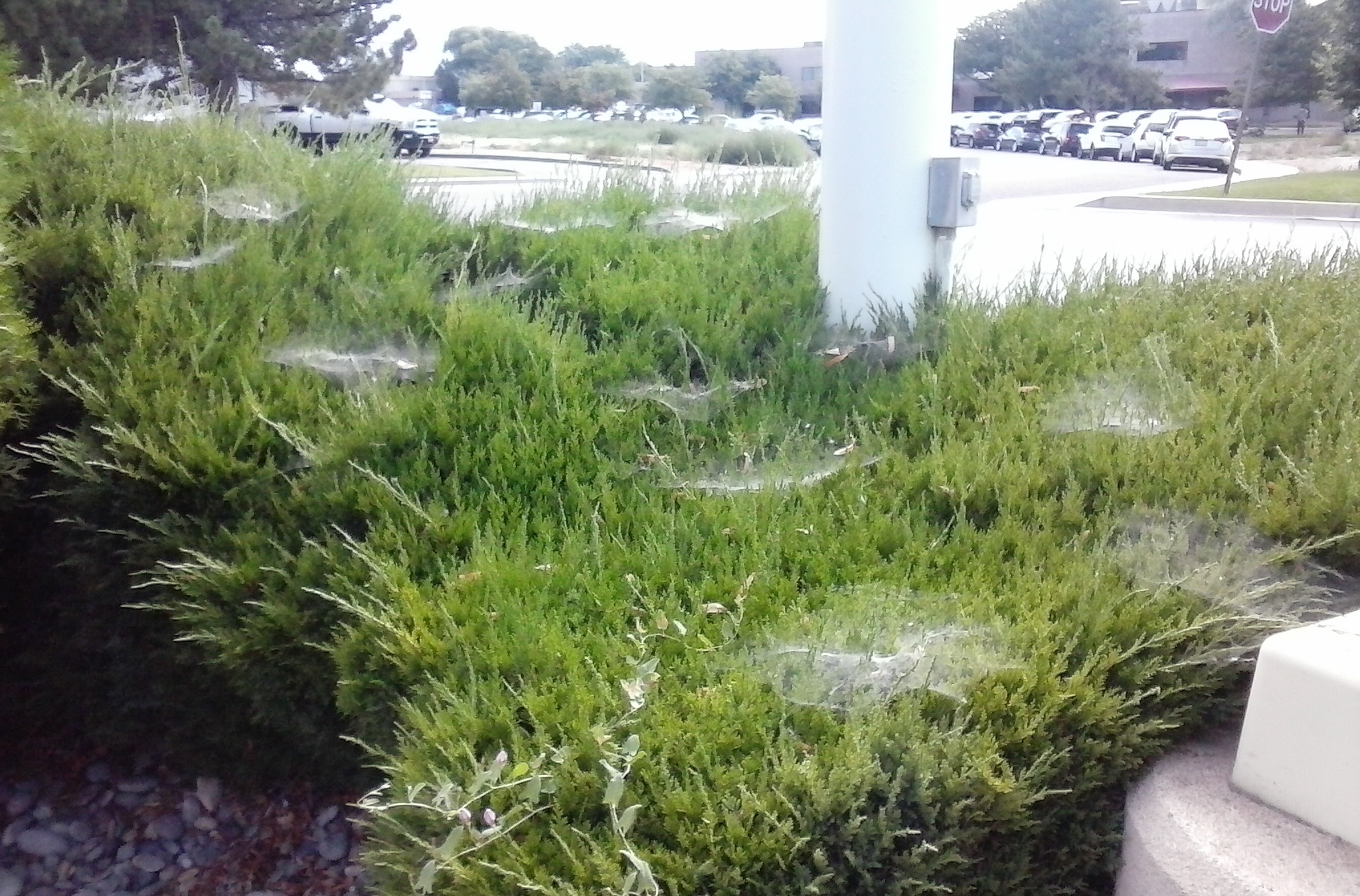
These things are, according to Steve Beddingfield, likely connected. And life-shatteringly important.
Since I didn't take a picture of when the 'blooming of white web-like stuff in the grass' occurred, and still occurs somewhat around houses in the 'greenbelt' in the neighborhood in the shadow of the Colorado National Monument, I can provide this picture, above, which I took the week students went back to school. I spotted this at a street corner in a business area near I-70 where it's catering to visitors / tourists. It's also near where the Rocky Mountain Health Plans office is and the interrelated Independent Physicians Association and electronic medical records / data businesses are. A hub for what was to be labeled by the power that be 'Obamacare' once the president was created to be the one to make that their 'lasting legacy' to the reform of the United States.
It is up to each one of us to take responsibility for deciding what kind of learners and researchers and providers of information, products and services we are going to utilize each step of the way on our path of living on Earth. As Lumigrate's logo / slogan states: Lumigrate - Lighting the Path to Health and Well-Being. You'll find tons of types of providers, models of medicine, schools of thought, possible products, books, podcasts, websites, YouTube videos, TED talks, and overall resources in the hundreds of threads at Lumigrate's forums. This is one, but it is one of the more unique and, I think, spot-on, pieces of information I offer at Lumigrate. I'm glad you've found it, I hope it at least inspires more divergent thinking and opening of the mind to see what might actually be in your environment and going on with the many things affecting you.
Who is Steve Beddingfield
Steve Beddingfield is a man from North Carolina who I encountered when he was out trying to engage new learners in a Lyme (Oregon) group on Facebook; the group had a strong leader and many very good members to learn with. He was then and still is, a 'household name' in a growing number of houses, certainly any I am in where the people are receptive to learning about THE Stuff (which is not always the case). Usually if there's a 'problem' people are more receptive IF they can see how the information applies to their problem or problems. Sometimes I can not find their 'spot', it's like on a dog the place it makes their back leg 'go'. Some are easier than others and sometimes, you get one you just canNOT find the spot.
His Protocol Basics / Overview
The Steve Beddingfield Protocol, I'll call it here, as it's sometimes referred to by his last name lately, but initially it was just called "Steve's Protocol", is given in full details at the extensive topic about his Facebook group, his protocol, his images and research. However, since this is a "101" introductory topic, I'm going to provide what Steve said in August to a man who appeared to be new in the group and asked what people were finding from being on the one man-made medicine in the protocol (fenbendazole); Steve added info as well about the Mother nature and man-produced medicine (red reishi mushroom):
Wild fungi are not safe, they contain the disease after it has been removed from the tree. Organically grown from sterile media is the safe way to obtain medicinial mushrooms. I cannot afford the tincture, so I go with capsules of Gano Ultra from Aloha Medicinals online. Half Hill Farm online has a great tincture.
Fenben: many sufferers respond to miniscule amounts. If one is heavily infested, its best to start low dosage with fenben. Reishi at first will start the bowel to purge.
This shows change in what Steve advises which demonstrates more experience. That experience up until spring of 2015 included some women who were of great help in the burgeoning group, at that time it was growing explosively in numbers. I was concerned because Steve would tell people to hop in with all parts of the protocol fully at once, and just figure out how to work around any side effects, things would work out, come out. Constipation is the primary issue with fenbendazole if there's a lot of 'stuff' in the intestines that become stunned from the medication's effects, which is how it is designed to work.
I personally had thought an approach would be wiser / better of taking the reishi initially, cleaning up the water source and food exposures, and taking the right amount of magnesium for a person to get their intestines working well enough, as constipation is a frequent symptom of chronically ill people before starting treatment and then adding the fenbendazole and starting with a very low amount and adding to it, adjusting downward on it and the reishi as needed. So here we are, on the same page in late summer of 2015 when I'm doing this overview topic. Glad to see it.
Here's the graphic produced by "PosterGal" in the spring, showing the protocol as recommended at that time, in one page without photos form, very helpful.

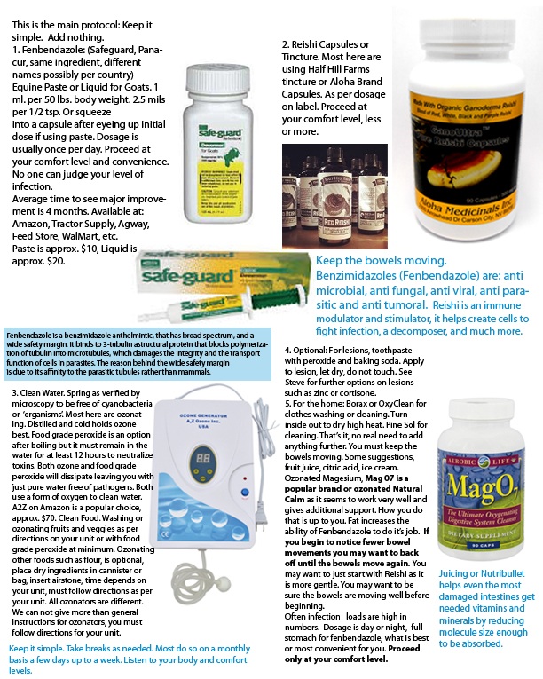
Please take the links in this topic to study the full information at Lumigrateabout Steve's Images, research, protocol.
To show how The Ripple Effect is working relative to Steve's work, here's a link to a website (titled How I Recovered dot come) created by a younger-middle aged man in the Boulder, Colorado area who has been working on reversing symptoms of 'tanked health', doing the usual things we learn about for environmental illness (heavy metals, nutrition, etc.) and then he was guided to Steve's information and created a nice overview about fenbendazole. howirecovered.com/fenbendazole-fever/
THIS HAS EVERYTHING TO DO WITH SAFE WATER, ITS IN THE WATER;
Ironically I Created SWIG in 2012 -- and in 2015 Steve's research findings made me more concerned than ever about safe water. Yes, it's great!
 Dr Jim Kennedy (DDS), Mardy Ross, Kevin King, Dr Jeff McCloskey (DC), August 2012, thanks to The Chateau at Two Rivers Winery for allowing us to use the backdrop without charge for our photograph.
I think we've all become aware that what we were taught by mainstream was not always the truth, or it was not fully making us capable citizens, at the least. Will the students going back to school learn about these important CURRENT EVENTS which have to do with THE Stuff? Will they learn about THE Stuff? As I edit this on the second day, there's now another story about tadpoles in many countries with a bacteria. Will they learn that today?
Will there be a concern about stories like this one, which ties into what this topic is about (fungus killing snakes, initially in the last weeks we were told one state, now it's at least nine):
abcnews.go.com/Technology/wireStory/mysterious-fungus-killing-snakes-states-32974960#.VclPDoCwzac.twitter Here's what you'll find with the social media share of it as a synopsis for our introductory purposes here:
"I think potentially this could overwhelm any conservation effort we could employ to try to protect this last remaining population," said Doug Blodgett, a biologist with the Vermont Department of Fish and Wildlife who has been studying the state's rattlesnake population for 15 years. "We don't have any control over it. It's just completely out there in the wild."

Hidden on hillsides in a remote part of western Vermont, a small number of venomous timber rattlesnakes slither among the rocks, but their isolation can't protect them from a mysterious fungus spreading across the eastern half of the...
ABCNEWS.GO.COM|BY ABC NEWS
There is a collection of stories on a comment thread on the Master's level topic about Steve's research, titled about mainstream telling us about this stuff. I'll leave the stories here just to those which were actually seen be me without any extra effort this week in creating this topic.
Same day as this was being shared by ABC News, this is what I found Steve Beddingfield out on Facebook 'getting word out about': Link he was referring to is: materread.wordpress.com/2011/07/07/we-dont-know-it-all-newly-discovered-deep-within-the-earth-worms-from-hell-hope-for-radiation-clean-up/
And what he has to say to explain is this, to a group of people focused on what's coming out of their skin lesions with a condition called Morgellons Disease, increasingly problematic in the US:Global warming caused upwelling of ocean currents, tropical storms picked it up and out if the salt marshes, then threw it upon us. I'm finding clams, mollusk so high up in the trees. Have pics!(Hurricanes) Andrew, Hugo, Sandy, and Katrina really got us good, she messed up our resevoirs with this bacteria, we now have polluted ponds and waterways, canals, then came the deep water oil gusher; BP GOT IT WRONG, MADE IT many times worse for us (with the things they did to remediate).A giant bloom of marine bacteria came here (North Carolina) in the clouds produced by the bloom. Having gotten caught in one while hiking, I can say for certain that this cloud was full of bacteria, the entire cloud was around me and it was a dark red. Have pics.
And so it goes. He beats the drums he feels he should, and I beat the drums I can. You'll see, below, the photo of what he found in a tree, after this event he refers to, perhaps recently. THE Stuff is all over the place, on everything, in everything, and has been making everything increasingly unwell. It has 'drained us', and for those of us with intestines, it's coating the small intestines along with a bunch of other stuff involved with it that we get inside of our 'gut' and making the 'microbiome' of the gut not be right. Not just for people, it comes in on our lettuces and the horses' grasses. And everything. It gets included in whatever is in the feed we buy for animals and then into their GI tracts AND it's in the water.
Are we treating the water correctly we're giving our selves, our other people, our pets, our livestock, our plants, indoor and outdoor, edible and decorative, and our fish? Ironically, the aquatics people I've talked to that take care of fish or sell stuff for indoor fish displays are more aware of THE Stuff than anyone. But they didn't know that it was something causing us to be preparing our foods differently. They WEREN'T THINKIN'. So that's why I say over and over, starting above, do what YOU need to do to THINK THIS THROUGH. TAKE THE TIME. MAKE THE TIME. THIS COULD BE THE MOST IMPORTANT THING TODAY AND YOU DIDN'T KNOW IT. I think it is. YOU might not. But at least look into this enough to decide what is right for YOU.
This was posted in a group that's focused about the increasingly-known disorder called Morgellons. Some will say we all have Morgellons, it's just that few of us know it SO FAR. I knew when I saw the symptoms it was one of the overlapping conditions, yet it wasn't listed with the overlapping conditions from the sources that so far had been 'all about the overlapping conditions' way of looking at complex chronic fatigue, pain, cognitive issues, etc.
This is what many of us think is needed to be learned about RIGHT NOW, shown in pictures, below. Actually, yesterday, we needed to know about it long before this, in my opion, if we were to be as well-prepared and strategized as those 'in the know' certainly are by now.
Since I worked in health education before completing my occupational therapy degree and doing that for 15 years, I've developed this analogy: Say you knew you were going to have sex with someone in the summer of 2015 and you knew you had a really tricky bacteria that the partner would be affected by BUT you were getting some treatments with chemicals that maybe were going to work --- would you tell the future sex partner before summer of 2015? It might be somewhat lessened by the chemicals you were being given and so, maybe it would be best to hope for the best and plan for the worst.
This ultimately is akin to what I think our government has done. I also suspect the Jade Helm training exercises being scheduled for July 15 and into whenever, a few months was the timeline I saw, had everything to do with 'this stuff' that I strongly suggest you TAKE THE TIME TO LOOK THOROUGHLY THROUGH THE INFORMATION WE ARE PREPARING HERE FOR YOUR benefit. This is the trough with the water, you're the horse. It's up to you. On with the pictures:
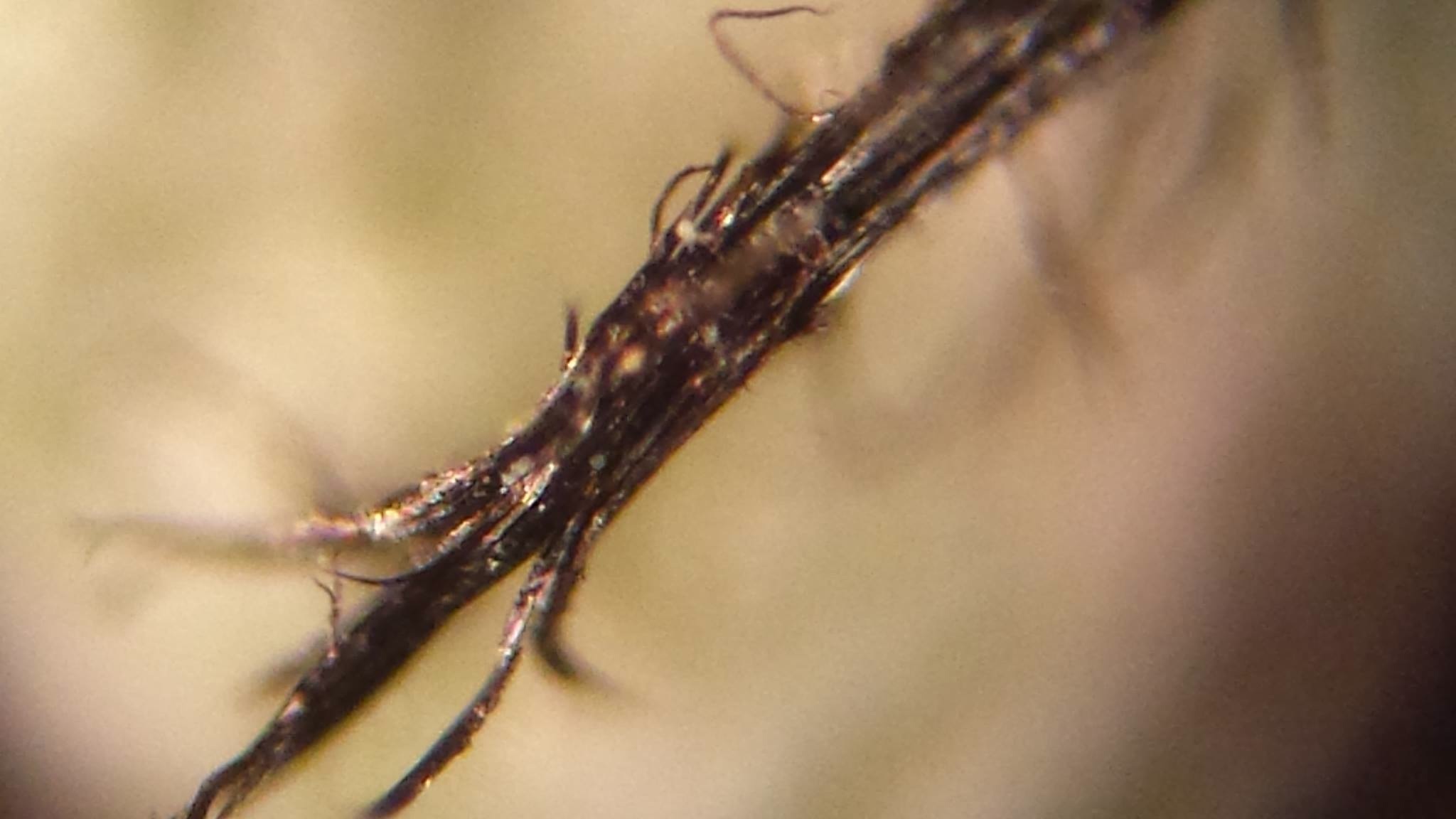
AND ........... 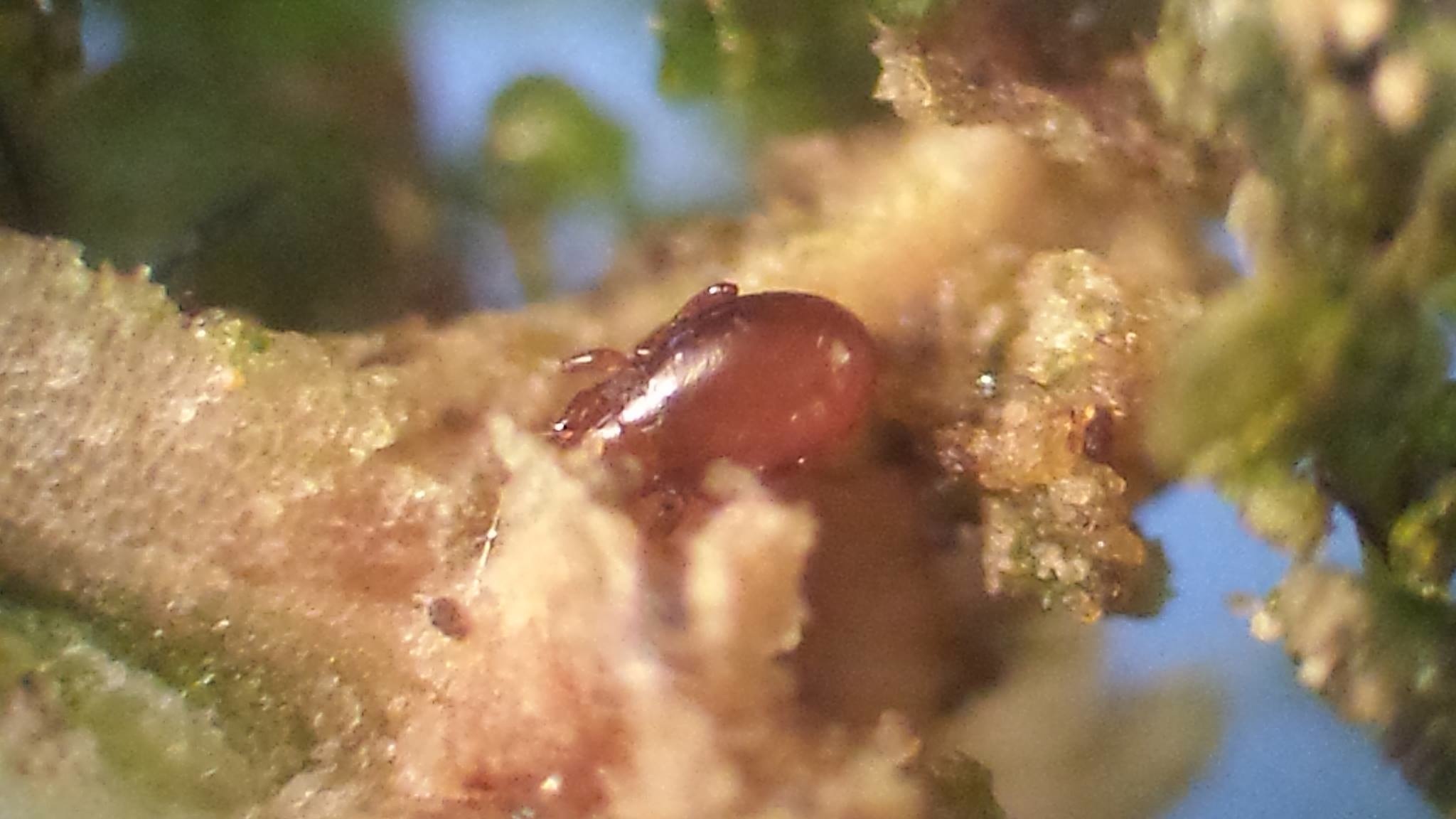
Both images, ©2015 Steve Beddingfield. Both 60X magnification.
TOP: GSB on a stick from a tree outside his home in rural North Carolina, outside Ashville, the GSB is the little white dots.
The other picture, above, is a "tick" clone sitting next to it's creator, GSB.
Level 101: Being created by GSB via it's horizontal gene transfer mechanism.
Level 201: All around the tick is GSB, its the cells, part algae, part GSB, NOTE THE WHITE AREA TURNS TO A GREEN AREA OF CELLS. Green has chlorophyll for energy; GSB uses the chlorophyll by growing a form that some call algae, but what is it when it's red and white?
I (being Steve Beddingfield) find red and white to be fascinating as a bio combo, but for now let's discuss green and white. GSB based "algae" is a round sphere with cells, a large central vacuole and replicates much like yeast. Both replicate by budding, it pinches off a new cell, a daughter cell, this leaves a bud scar at the thermal point. I'll get some pics, and be back to place them here. --- This is an example of what it's like to be in Steve's 'classroom'/Facebook group.
GSB as a "Trojan Horse" for algae, is how he titled/explained this picture, also ©2015 Steve Beddingfield.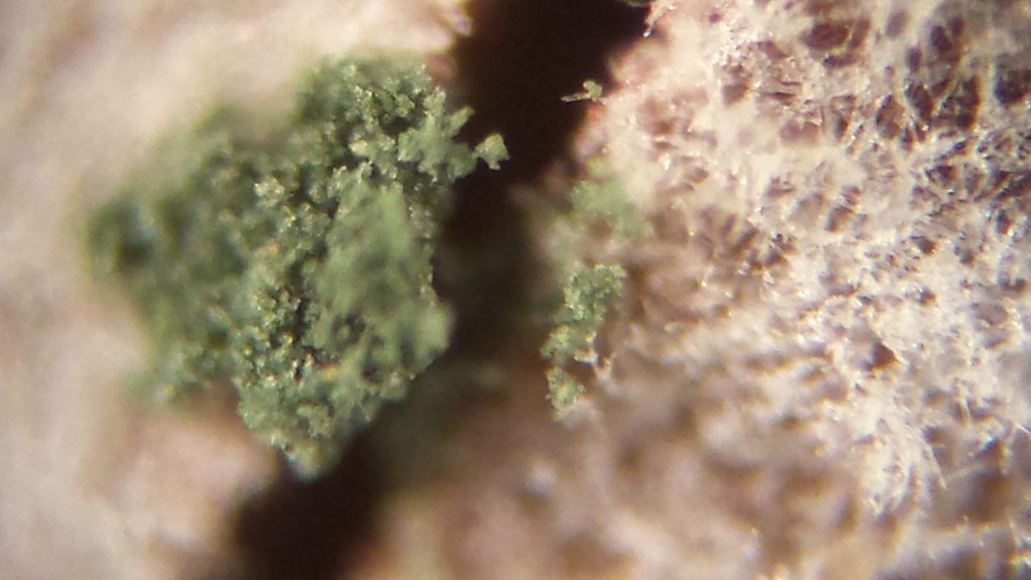
"The spores of GSB, they are performing lateral gene transfer. You have permission to use these pics for analysis and referral."
© 2015 Steve Beddingfield
Does THAT get your attention? I hope so. Please consider taking the TIME and making this a priority to pour over what is presented at this topic on Lumigrate AND go to the "Master's Level" topic if you get this one under your belt.
It has a lot of facets, it will take time to digest, so go away, come back, go on, keep at it. Digest. Form opinions. Decide what YOU think, and then, as always, be proactive/ take action if you so desire.
Here's the link: www.lumigrate.com/forum/steve-his-images-his-protocol-and-facebook-group-community-and-other-resources-chronic-illness
When I got my "head in the game", Summer 2013, When This Occurred.
My ankle, August 5, 2013. Those spots were on the tendon above my right ankle on the 'outside'. It was right where the hem of my jeans had been, and occurred just after I'd been outside in the greenbelt near my home where I'd 'noticed' previously that there were tons of what I mistook to be 'spider webs' in the grass. The grass in the summer of 2013 was overwatered, my feet were splashing around sometimes it was so wet.
Yet the grass wasn't looking very good. I was intently taking photos of chemtrailing, which had started on a massive and daily basis on June 1, 2013, about 6 pm or so. We had our first 'hammering day' where there were always several aircraft spraying in the sky all day long, on July 31, 2013 and that was when I got the exposure, being outside for hours that evening taking pictures, posting on Facebook and being an 'activist', teacher, learner about whatever this geoengineering stuff was about!
I didn't know, but I knew it was important to cover for health and that's my 'beat'. The grasses this year, and the trees, look much better. It all makes sense what I witnessed and couldn't figure out before connecting with Steve, and what he has said.
The chemicals in chemtrails naturally are clearly known to not be good for us and contribute to health problems BUT the GSB trumps that in importance, it had to be and still has to be dealt with and reduced. Is there a way to eliminate it? I doubt that. So our due diligence in learning how it affects us, and how to work around it, reduce it, boost up our wellness of mind, body and spirit and DEAL with all this, is what this topic is ALL ABOUT!
There was an oddball scar for a while, it looked like a tatoo. It was visible to me this year earlier but not now, two years later. I guess I'm healing. Healthy enough. And I'm here to work to get the information to YOU. Thanks to Poster Gal and her crediting the truth tellers. Back at cha! And naturally, praises and encouragement for people who can do PayPal to get Steve's work going forward funded. This is what he said today: "My email is best for donate, goes to PayPal so no charging me or them. stevebedingfield@yahoo.com"
There's a lot I put in forums about chemtrails and geoengineering. Look if you're interested, I'm not going to suggest people take much time at this point to study that in depth if you've not already, I'd stick with this and going on to see what to do in your home for chems that will help on it, as it's all about doing things differently cleaning and preparing foods, and drinks and ice and etc. Oh, and not going out barefoot in the hard with pants that will absorb what's in the grass then rub it into your skin along a tendon when you already have funny skin from the EDS-type symptoms of 'this stuff we all have' as I have.
Now, I want to provide a picture that I took, of something I 'noticed' in Grand Junction, Colorado in the summer of perhaps 2012: This, Steve has said, is what to look for in the grass because it's GSB running amok and the exposure from consuming the grass (or hay), or otherwise getting exposed would cause problems.
We already have chickens in crisis in our food supply this month in the US, when will it be cattle? Hence something YOU can do is KNOW THE SOURCE OF YOUR FOOD AND TEACH THEM ABOUT THIS AND CHECK INTO THE SOURCE OF THE FOOD ANIMALS WERE FED. See how YOU can do a LOT about your exposures? You'll learn, below, about the simple (which is a relative term) steps to getting safe or safer water, and fruits and vegetable remediation. Again, best to have the producers doing it too, but in-home ultimately is where YOU can have the most impact the easiest. Most bang for your buck. Etc.
Lumigrate has always been about "lighting the path to health and well-being", as our logo shows, and the Lumigrate YOU! Model -- YOU learning to get the information and providers of info, products, services surrounding you (or those you are guardian of or helping advise). Again, the YOU! Model.
"Who Would You Add?" in the 11:00 position might make you think about someone like me, a layperson who has a background of 15 years in allopathic medicine (as an occupational therapist), a bachelors of science degree, on top of two years of experience at the student health center's health education department at Colorado State University. Today an information concierge, tutor, advisor, consultant about where to look for information, where to go for products and what do DO in your home, office, car, etc.....
OR Steve Beddingfield, who you'll be learning about here, extensively (and how to donate to his efforts).
OR "PosterGal" whose real name we leave you to be curious about, and since she makes frequent comments such as this one on Facebook, which is from early August 2015, I felt was best left to disclose her identity as PosterGal as she wishes within her circles. The problem is, she'd be an excellent advisor to 'hire' but she's dealing with this stuff as best she can having been very ill before finding the information and with a child and family to look out for. At a critically stressful time as things 'bloom' and perhaps implode for some or overall.
You might find your MD or compounding pharmacist or PT or yoga teacher is acting strangely, or is not well in other ways. Maybe they no show, or cancel, or aren't open. Little by little around you, there might be signs of the dysfunction. To me it's like when a hail storm starts and it's a ping and a pop and then a bam, bam, bam.bam.bam....... I've seen the frequency and magnitude of people's health issues explode recently.
Of most concern to me is that I've seen people's breathing stop. Others have said they've seen it with people. So I want to introduce here the concept that PosterGal turned me onto only recently, SIBO (Small Intestine Bacteria Overgrowth), and the concept of " .... hydrogen sulfide gas, a byproduct of sulfate-reducing bacteria which is released in the intestines of those suffering from SIBO, colitis, IBS and other chronic inflammatory bowel diseases."
Basically this stuff in us is making quite a mix in our intestines, which creates gas, bloating, and then elimination 'irregularities'. That's a chemical mix in there. Add something else to it and it can have different and more severe effects. Falling asleep suddenly, stopping breathing. Dying. This, again is the OVERVIEW, This Stuff: 101 topic, so I'm going to simply set up the link here to find the information on the "Master's Level" thread about SIBO, bloating, and etc. I strongly recommend if you have bloating, get a distended belly, have IBS, etc., you take this very seriously AT THIS TIME. You've gotten away with it in the past, but currently you can basically just get pushed over the edge. www.lumigrate.com/forum/steve-his-images-his-protocol-and-facebook-group-community-and-other-resources-chronic-illness#comment-2876
This is massive. Everyone's basically with their realities to live out and in many cases that is going to be devastating health outcomes and deaths.
The whole system is going to be stressed and undergo enormous changes and the NEW ways which are firmly rooted now will end up replacing them.
I'm hoping to take some time off. I've spent years researching. I thank the truth tellers for all they do but it's getting very hard to cope with, the scale of this devastation in our environment and how it is affecting real people and having to watch it everyday.
I know it like the back of my hand, these myriads of symptoms, I can predict and tell you what is likely happening. I'm not bragging, maybe it's some kind of talent, I don't know, or just having seen it and read it ten million times in everyone that writes to me for help, just experience.
The sick Mom's, the sick Dads, the sick kids, the sick pets. Maybe I'm just good at connecting dots, a good detective, I don't know. A doctor in the old days would have done much the same instead of relying on labs.
I know a few autism moms can do the same, spot them in two minutes. I need to disengage for my own well being. I am not going to save them all, that is clear..... so I will just have to prepare my own self and mind for what I know is coming. (Posted August 7, 2015)
This, below, from August 10, 2015, in my Facebook feed when going to grab from Facebook what I saw PosterGal had written about her state of mind, from a woman who had a most interesting and complex background, which included being displaced by Katrina with massive exposures and then she migrated north a ways to re-establish her life in the rural parts of Arkansas: She had provided a stunning piece on Lumigrate when asked, in recent years (an artist, a teacher, a deep thinker who had been hit by a train and had a significant neurological injury from that trauma on top of the environmental exposures....)
Drama Alert (as what i am about to write will be undoubtedly so translated by more than a few folk and by those of you have the need to categorize my actions while distancing yourself).
So in 5-4-3-2-1 days, I am deactivating my account. I need to spend time with me and use my limited energy in the hope that my life becomes once again meaningful and worth living o me. Tis a bad stretch of memories heading my way. And I must decide whether or not my decision to refuse to remain dead is the decision that best served my living. I am most weary.
I bring information to Lumigrate's YOUsers from an "integrative medicine" standpoint (mind, body, spirit or mental/behavioral, physical, spiritual it might be said), as well as "functional medicine" (basically meaning treat symptoms as minimally as possible and spend your efforts looking for the UNDERLYING reasons for wellness or illness and be active treating at that level. Which brings us to this graphic, as I was wanting to show how we'd 'peel the onion' of the layers and get through the many layers to the core, eventually (given enough time, energy, money it is possible to do so!).
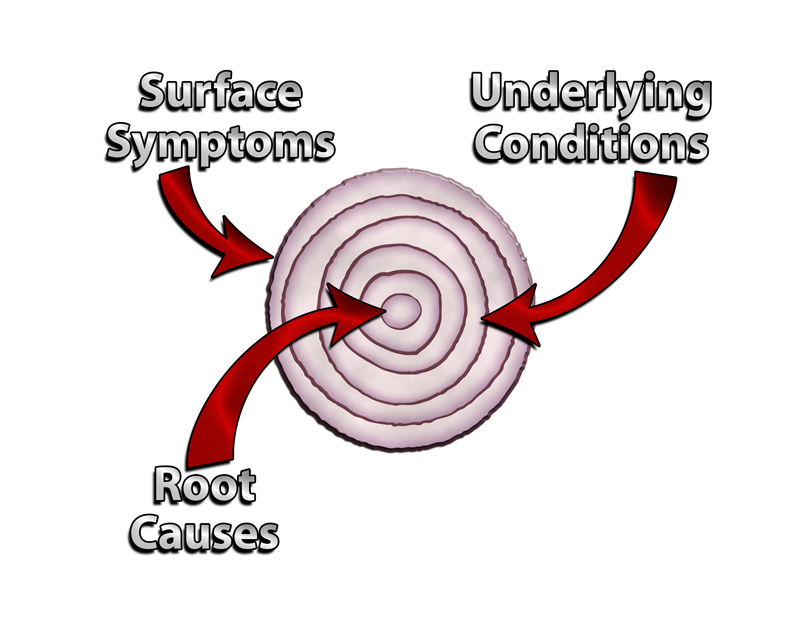 With the information I'm presenting here, stemming from Steve Beddingfield's research he's provided, I'd suspect he'd say the root causes can be singular, it's giant sulphur bacteria, or GSB. The underlying conditions can be the various disorders caused by the GSB 'collaborating' with the alpha organism it is symbiotic with, which varies.
This is why, after what we think is an algae bloom (which is an "algae" bloom, we'd say in quotes, as algae is now a hybrid or morphed organism with the GSB, so it's not algae, it's "algae") you can see up to 25X (25 times) the number of cases of a certain identified, category of diseases, such as ALS or MS; but you won't see a mix of both (unless there were two things in the area that were the alpha organism the GSB got 'going with' replicating.
If the bacteria that causes Lyme is the alpha organism, it wouldn't be Lyme, but "Lyme". Mold is now "mold", and so on and so forth. The outward symptoms on the surface are the lesions on our skin, or red bumps, white marks, brown patches, cysts thought of as acne, joint swelling, pain, stiffness, aches in the ears, the head, ringing in the ears, floaters in the eyes, laxity of the tissues of the body including joints, and so on and so forth.
This thinking fits right in with our long-standing "load theory" model, demonstrated here with an elevator which starts straining and running with symptoms telling us it could break and crash soon, due to the 'load'. So think about all the things 'on your plate', 'on your elevator' that are loading you down, offset by things YOU do to boost your wellness up --- and i
If you're doing a bunch of things to help your wellness and were unaware of the GSB, you might have been doing well with your wellness level, relatively speaking. But as the GSB gets to doing it's thing where it gets into you (through foods and drinks, the water you shower/bathe in, breathe in as vaporous steam, etc.) even those being proactive might have symptoms of unwellness increasing. Become AWARE of this information AND LEARN IT enough to be correctly proactive (correctly being a key, operative word), and you clearly reduce your load again. That's ALL that needs to be done.
However, if you've not been proactive in ways that have been really benefitting your wellness and you're currently with a problematic wellness/illness level at the time you learn this information, you're going to have a challenging road ahead moreso than the others who are 'weller'. Or if you're seeing this occurring, as I am, in people around me as I've shared above with YOUsers reading this topic, this might help explain why people are going to come to pass. Either from our Facebook because they're turning to their live worlds to deal and clearly possibly die, or just to put their energies to what will ultimately help them (and their families, homes) the most.
This has EVERYTHING to do with 'spirit', by the way. What people believe via their spiritual belief systems is having a drastic impact, as always, on how they interpret what's going on around them. Please be aware of this and use your wisdom about it. That is all I'm going to say here about spirit. (Additionally, we have a spirituality coach on Lumigrate, go to the About tab to see how to contact me and I can connect you if you are appropriate for Beth Patterson and vice versa.)
Behavior/mind, we see this also being affected. The protocols for treating autism often go by the moon cycle and there are reasons for that --- the 'interlopers' inside us that are contributors to the symptoms are affected by the moon. So this is worthy of note; the sicker someone is in the gut the more likely they are to be sick in the mind too in one way or another because it's this simple: the brain is part of the body.
(Additionally, we have professionals I am linking to from Lumigrate to help with that, go to the About tab to see how to contact me and I can connect you if you are appropriate for Beth Patterson and vice versa. We're not a crisis center, however, use the appropriate mechanisms in your community and many do not know to look up their county name and 'crisis line' and that's a good resource typically. 911 will also get you there. My focus and niche is getting to as early intervention with education as possible and helping people implement things in their daily lives.)
So there's a lot of melding of "mind" and "brain", it's a grey area -- no pun intended. Grins.  but for our purposes here, the effects from the interlopers on behavior are actually body not mind. Mind is more like 'mind over matter' -- we can work really hard to keep our mental attitude, despite what is going on in the body, where it benefits us. Or where it harms us more. And that's all I'm going to say really about that here.
Let's focus on the bacteria, the interloping microbes, the interacting symbiots, and the things that Steve Beddingfield has been able to take pictures of. (Symbiotic means you help each other out, basically. Synergy or synergistic means the sum ends up being more than it would be if it were straight addition. 1 and 1 is greater than 2, in other words.... just a little vocabulary help here, up front). You'll see the word 'symbiont' referring to things in the photos.
I have a sense that this fall semester that is underway now, starting in the days where we're well into the long days of summer, after the whole wet and warm season has had it's time to 'do it's thing' is going to have things happen that is 'history in the making' which will be affecting greatly the course of the future of our nation and beyond.
And we are not alone, naturally, our governments around the world, and therefore the people, will all be affected one way or another. Mother Earth is getting a cleansing. And it is going to be a challenging time like nothing we've ever seen and perhaps our elders and those who were here and told us about the Great Depression, the Dust Bowl, World Wars. Our attitudes and our energies have everything to do with affecting, collectively and individually, our relationship with what occurs.
This is going to be the simplest, most straightforward topic I've ever created at Lumigrate that is of significance. That's because it is 'time of the essence' and has to be appealing to as many people as possible, and present enough information to show people how important it is. I don't have a 'crystal ball', but what I do have is a lot of autism-type characteristics, similar to my father who passed in 2010 and cared enough about me and my work to have supported it in the capacity he could. I also was gifted to inherit much from my mother in things she had similar abilities with, forseeing the future, fortelling things and this is, unfortunately something many religions teach people to turn away from and cast out those who are known to have the abilities. I'd be lying if I didn't reveal it, and I was reminded recently by a woman I felt a strong connection to, that this is the case. Turn back now if you wish, I'm just being honest about where I'm coming from. Proceed if you wish, and I believe you'll benefit no matter what your personal belief system is spiritually.
What my mother and father provided to me upon their passing, financially, allowed me the time to be working literally daily on information to load into the troves at the forums tab at Lumigrate.com. So be prepared to be linked IF you want to go into more details, to select topics in the troves at the forums tab. Just as those funds were running out, I connected with the information source, Steve Beddingfield, I'm collaborating with to bring this information your way.
From within his group on Facebook, I then connected with others, who have 'spun off' since then, so you see, there's 'the ripple effect' going on, literally before my eyes! It's wonderful to see! However, it initially presents as a 'divorce' or 'breakup', 'fracture' might be another word.
It's similar to what I experienced which really broke my heart in the first years of Lumigrate (which launched in spring 2009) when the integrative health center I'd help get off the ground similarly fractured. The advantage of age is that you have the years to move forward and see backwards and realize that we're all here still --- we've all served people, just differently, and it was essentially a ripple effect that benefitted the consumers in the public sector we serve more than had we all stayed with our initial intactness.
I admit, I dismissed the images posted by Steve in 2014 that went by in my Facebook feed, as it was mostly over my head, or not being taught in a way I could understand. Pictures of things he was seeing in his microscope and teaching to 'autism moms' or people with complex chronic conditions OR BOTH. It was on Christmas Eve I saw a gal that was BOTH (diagnosis MS for mom, daughter with autism) posting about going to Mass, and 24 hours later she was swearing like a sailor, angry about the medical system. That caught my attention and drew me in. Later to be nicknamed by me PosterGal.
Nobody at this level of learning and proactivity as she is at would be wasting their energies and others' on complaining about insurance-based, organized, allopathic, 'modern' medicine, we all have long-ago figured out the monkeybusiness it's about. What was she angry about?
She'd found what I call 'The tell-tale patent trail' (that you can get elaborate detail on if you wish in the subsequent information I provide later at this topic). Or simple The Patent Trail. Just as The Ohio State University is so important they added 'The' and I am told are known in their circles simply as 'The', I point out 'The' in this title I've bestowed upon her findings. Up there with other life-changing moments, I will not forget the feeling I had, the thoughts I had at the moment that I realized I was literally with a front row seat to medical history in the making, it appeared, within Steve Beddingfield's group with PosterGal having just found that patent trail.
I eventually went through to see who'd been added the group before me, and most of the gals who helped Steve's work blossom had been added before I had, but not all. Credit to them all for what they did. I asked for interviews of the main ones, and they all just started talking to me but it was launching to a spiritual calling they felt they had for helping assimilate or educate with the information.
Then the 'split' happened, and I've simply wanted to recognize EVERYONE who has contributed to the process, as I hope they will fairly recognize my participation too. In making this topic be the best it can be, I'm going to be approaching both 'parents' as the 'child' of 'divorced parents' who have respect for each other but who have broken up.
But my place is for bringing YOUsers the best informat I can, and to do that, it means asking for input from others and so I will be asking for input from Steve and PosterGal and potentially others of the 'et al. gals' who knew the information so well, were advising others so well, and spun off to create new groups and be independent on their walls helping people learn. As you see, above, feeling the burdens often, too.
And despite my being in a major place in my life overall, at Christmas 2014, I found the time to communicate with PosterGal (and the others). I told her of my greatest 10# teacher, my cat who I'd had to euthanize in 2013, who had what clearly was sensory integration dysfunction symptoms and I thought, autism symptoms. "PosterGal" as I call her on the Internet, told me to look back in the Facebook group to something she posted about a month before about horses and this stuff that we're here to tell you about in this easy-to-figure topic.
She had posted a deluge of information once her vision returned, her energy was increasing and she was quite frankly, REALLY (______) .... mad. She revealed to me she'd gotten into the research full force because she was 'contrary' in a sense, she was wanting to prove Steve Beddingfield's work wrong because it was so simple, his protocol, and inexpensive and that meant that she'd just spent a lot of money, time, energy in the past on things that were not the 'real deal'.
The anger I was drawn in by seeing expressed on Christmas afternoon was to do with this: if a layman like Steve could figure it out with the help of an autism mom graphics artist like her, and other women (and men in the group too, to a lesser extent) who hopped in and helped prove things out in 2014, then those who had gone to college, who had shingles hung out providing to patients in return for funding from them should have as well.
But I had to dig back a long way to find the article she was telling me privately to look for in the group about horses that seemed to be describing what in a human is called autism. As long as I had to dig back, I may as well organize the mess of links into categories of the protocol and supportive information, and so I did that. And I created a topic as soon as a could in early January, on Lumigrate. And it's had more reads overall so far than any single topic ever. I've always let the read numbers help guide me, so the numbers and my gut were in agreement!
I daily would participate in Steve's Images group on Facebook and pull out the highlights that I saw happen there, and change and improve the topic on Lumigrate. It became very long and detailed, and is a wonderful resource. So this topic is the 'primer'. Think of this topic as "The Stuff: 101" and that first, more complex thread as "The Stuff: 501" -- Master's Level. Sometime we'll maybe have a doctorate level version, specifically for doctors of all kinds.
I'd see, in early summer, a video that further confirmed my thinking about autism in humans and other animals being the reality; from another 'autism mom', nicknamed 'The Worm Whisperer' today, whose focus dropping into her presentation at a conference this year was about the older more complex children with autism who become violent. And the worms they have in them. And the symptoms that improve when they get treated.
Her passionate, emotional story about how he attacked when having an episode reminded me of the way my sweet cat had attacked me, in what was known to be a brain 'miswiring', and seizure-like. Her pupils fully dilated in these episodes; PosterGal's child had that symptom when her emotional meltdowns would happen. My 'autism-like strength' of collecting dots and connecting them, and my mind not resting until it did, was 'doing what it does best.' I felt I could be part of this history-in-the-making, and now had the reach of Lumigrate as a platform to offer what I could.
Just last month I had time to do a little work again on feline autism information and there was finally a DVM in the USA who had posted a video at her website about feline autism symptoms responding via her holistic, homeopathic treatments. She'd simply known enough about autism in people, to make the connection with a customer's one cat. And was I surprised when I saw the cat and it was also a 'tortie' like my beloved '10# teacher'? No. Two cases of it were in the national news when the owners called 911 when viciously attacked when arriving home, which is what happened in my first attack in 2012 as well.
SO, we've introduced kids, parents, other adults, horses and cats. Dogs are not exempt: As a matter of fact, dogs are in the news more than anything. Stories on dogs dying of cyanobacteria after getting into lakes or other water areas that have been posted to forewarn about algae blooming and being unsafe. They'll sometimes rapidly die, if the intervention isn't fast enough, correct tenough, etc. But why wouldn't it be posted to forewarn about dogs? Why aren't ALL dogs dying, hence people more aware? Are people dying? I believe so, but 'we' might not be attributing it correctly. Will more people die soon? I think so.
What about chickens? I think the story about the recent egg shortage and increase in prices, debacle over so many chickens having to be killed or dying that disposal is a problem made it into the minds of more Americans than any story I've seen that has to do with 'this stuff'. Why was it the factor farm chickens and everyone around Grand Junction who has chickens in the yards are not concerned? Because it has everything to do with WHAT IS IN THE GUT / INTESTINES of the animal. And they'll be more or less affected, too.
Have you seen animals lately acting oddly? I have. Rabbits laying down in the middle of the road, playing dead. Coyotes with plenty of rabbits to eat getting aggressive with dogs walking with owners.
In humans, the vagus nerve is 'the superhighway' for things to get from the gut past the blood brain barier and into the brain. Ever notice there's not a single person anymore who doesn't have an endocrine problem, a mood problem, a behavior problem, an attention problem, a spatial orientation problem. Let's talk about that for a minute. Research allegedly stumbled upon by a major research university revealed recently that brains of sea mammals that get beached have what researchers believe to be cyanobacteria in them. Same with human brains of those with attention and spatial problems (think 'dyslexia', or 'I don't know west from east, tell me the directions another way').
And This is How It Happens
Teaching is not easy; it's the reason teachers have students start teaching the class early on so that they get an understanding of what it's like to stand up in front of a room full of people not looking like they care, 'get it', etc. And then we all get to have fear of public speaking until we 'work on that'. HA!
There are a lot of different learning styles, even 'ideally', when everyone's brains are working well it's complicated enough to provide the various kinds of learning. How many brains today have the wellness to be able to do the work involved with complex learning? The education system has lowered the bar as a result, or they'd not have enough students in seats to pay the bills.
It's almost a case, with 'this stuff' we're teaching here, of 'which came first, the chicken or the egg?', so be prepared for that, I suggest. It can be very complex. But this version is intended to be very simplified.
The Earth has a lot of places where things come up to the surface from below, carrying with it what is down below. Over most of the Earth there is the ocean waters, hiding from our view what is going on, but we have explored the depths of the oceans and some of us have had the ability to see what others capture there, and some are lucky enough to see it first hand. I should have known something was up when two of my friends from growing up in the Colorado mountains, who raised their families in the area, had daughters who went into marine sciences; the oceans are 'where it's at now'.
Coming up from below into the cold depths of the ocean were bacteria which have done the most incredible things. Around the Earth we all could see the space, the stars and always long for exploration and wonderment out there, and that's the direction much of our funding and exploration went. But ultimately what is going on at the floor of the oceans is what 'everyone is rushing to explore ... and some to exploit'. Solutions in medicines, ores such as gold and iron, and many things are perhaps being warred over without our awareness.
This potentially might include those who would be called 'aliens', and I'll simply defer that by suggesting people might consider watching what History is showing on the Ancient Aliens program. Not considered of overall value by many thinkers, I've reviewed tens of shows now and find that in each one there is substantial information which I've seen substantiated in various ways. I would at least consider it a source to put time into if one has time to put into such things.
For those who want my fast overview, they gave a very good overview in 2011 of what Morgellons disease is caused by, which satisfied even the more critical Morgellons activists from my asking around. And the season opener for 2015, in July, told us that a few years ago bacteria was found on a very old space rock. Another expert they interviewed elaborated on how it is that the cold and hot and ultraviolet could be dealt with by the bacteria when in space. Had I not been workoing on Steve Beddingfield's information for a year at that point, intially only peripherally paying attention and more recently daily dialed in to learn more, I wouldn't have known the significance of that segment.
I now say that I consider the Ancient Aliens program a 'pace car' of the disclosure to us of the information which Steve's research is focused upon. And what is the core element in Steve's research? Giant sulphur bacteria, or GSB. Basically, GSB does horizontal gene mutation with whatever it gets onto, which I will call the 'alpha organism'. This can be a bacteria. A virus. A fungus. An insect. A -----
These bacteria, naturally, have always had a 'job'. They clean up things ... they 'feed on' petroleum (or today products man makes that are made from petroleum such as the massive amounts of plastics we have out there in the environment). They also use as a source of fuel phosphates. So just stop and think about the history of modern man and our increased use of these things that got into the environment and daily continue to be added to the environment.
So they have always been hanging around at the bottom of the seas, have always been getting airborn and then transported and coming down onto the soil, onto the snow, onto the ice and glaciers, has had a LOT of changes lately. Super storms such as Katrina and the massive tornados have stirred things up, making more than ever go airborn! And when they come down to land after they've been transported in whichever ways, they'll be thriving if they have MORE things to feed off of than less. So think about what parts of the country and world will have more or less problems. Add to that what we know about heat and humidity factoring into things 'growing'.
We all know, I hope, about the overall, massive melting of ice (such as glaciers) and snows banked; there's a red ice shelf in Antarctica that's thought to be 'cyanobacteria' but, as you learn from knowing 'this stuff, it's actually cyanobacteria symbiotically having 'relations' with giant sulphur bacteria.
The information studied and published even for common people to get hands on which is on what is thought to be cyanobacteria and about every condition there is labeled from A to Z is extensive. That is included at the "This Stuff: 501" version of the information for those who want to study more if you feel you're read to go deeper and MASTER the information.
Steve Beddingfield's information he was teaching in the fall of 2014 was focused on cyanobacteria; the first I saw him mention GSB was late November 2014 and it was around Thanksgiving, so maybe he was just being a turkey. Nobody much commented about it. And I didn't see reference again by him or anyone to GSB until one day in the spring he basically dropped the 'cyano' ball and picked up the 'GSB ball'.
But this was the timeframe I was trying to catch up on learning, creating the 'Master Level Topic' YOUsers of Lumigrate have to explore, and so I was barely caught up to understanding the information when that happened. I was again confused. And now I hope I'm demonstrating my competence with this information in how I'm presenting it. I will be asking for feedback on it from those in the know who I refer to hear as those who help me learn. I hope by my admission of the difficulty of this information for me, it encourages YOU to keep at it.
A key element in the process for me was when PosterGal found The Patent Trail; the patents are for chemicals similar to the chemical fenbendazole, which is what Steve's protocol for treating 'this stuff' includes, in addition to red reishi mushroom extract and purifying water, produce, the air, how to clean your clothes, sheets, towels, environmental air and homes, cars, etc. This Stuff is a real 'game changer' and of utter importance to have awareness level accomplished. Act upon by learning and becoming competent. Change your lifestyle and activities to reduce exposures and to consider doing the protocol or components of it. I suggest everyone start with cleaning up their water and how they treat their produce. Everyone. ASAP, now.
And then next I'd suggest making a decision about red reishi extract. And then beyond that would be thefenbendazole, which freaks people out because it's a veterinary medicine. It's over the counter, used for a long time in many applications. The patent trail for the other chemicals like fenbendazole, not yet developed to be on the market that we are aware of at least, is where I'd suggest people learn about THAT element to become comfortable. Their supportive documentation cited an incredible gamut and array of disorders that were proven to be affected by the chemicals in the patent requests. THAT IS WHEN I WAS SURE THIS WAS HISTORY IN THE MAKING, but YOU have to decide for YOU. (And those you make decisions for.)
We've seen what the 'autism' community is saying about parasites, what experts like Deitrich Klinghardt has said about parasites more recently; I had been studying that, providing THAT at Lumigrate, prior to encountering Steve Beddingfield, so my 'path' had been lighted already. (Lumigrate's motto of "Lighting the Path to Health and Well-Being" what I'm referring to here). So I have that to link to for YOU for further study, as you go past mastering This Stuff: 101 to the 201, 301, 401 levels, with many of my links taking you to the thread that I would consider giving you the Master's Level 501 information.
BUT THESE EXPERTS have, to our awareness so far, not gotten ahold of the information that Steve Beddingfield presented to us, offered at Lumigrate's topics for the highlights. However, I have repeatedly said to his group and others, Klinghardt in 2014 did an interview on One Radio Network with Patrick Timpone where he said that chemtrails and geoengineering is the biggest story of his generation. He in the same breath practically said that 'doctors know a lot and we don't have first amendment rights to free speech, we have commercial rights' and implied they were being censored.
Steve Beddingfield has implied and then stated more clearly in his group that his images and videos and what he is allowed to put out for others to see are also being censored. I don't want to cross that line with what I put on Lumigrate or on Facebook because they can mess with your website and many other monkeybusiness things. PLEASE people, understand how the monkeybusiness aspect works, so you can then value the information you see and know better how to navigate. I do feel that I'm staying within the boundaries of what the power that be will accept from Lumigrate, since we have such a relatively small 'reach' to the public.
Their concern is valid and I support it; people have to become aware in a systematic way so that we have enough people who can baffle the potential hysteria that will occur when this comes to be known by the MASSES. PLEASE take this into consideration when sharing this information to those YOU care about, who you influence and who are in your reach. We want the ripple effect, and it will become a tsunami, most likely IF I understand the implications and forsee the future correctly. But a well-orchestrated people can have a tsunami come ashore and it's much less devastating than when they're not well-orchestrated.
Live and learn. Learn and live better! ~ Mardy
__________________
Live and Learn. Learn and Live Better! is my motto. I'm Mardy Ross, and I founded Lumigrate in 2008 after a career as an occupational therapist with a background in health education and environmental research program administration. Today I function as the desk clerk for short questions people have, as well as 'concierge' services offered for those who want a thorough exploration of their health history and direction to resources likely to progress their health according to their goals. Contact Us comes to me, so please do if you have questions or comments. Lumigrate is "Lighting the Path to Health and Well-Being" for increasing numbers of people. Follow us on social networking sites such as: Twitter: http://twitter.com/lumigrate and Facebook. (There is my personal page and several Lumigrate pages. For those interested in "groovy" local education and networking for those uniquely talented LumiGRATE experts located in my own back yard, "LumiGRATE Groove of the Grand Valley" is a Facebook page to join. (Many who have joined are beyond our area but like to see the Groovy information! We not only have FUN, we are learning about other providers we can be referring patients to and 'wearing a groove' to each other's doors -- or websites/home offices!) By covering some of the things we do, including case examples, it reinforces the concepts at Lumigrate.com as well as making YOU feel that you're part of a community. Which you ARE at Lumigrate!
This forum is provided to allow members of Lumigrate to share information and ideas. Any recommendations made by forum members regarding medical treatments, medications, or procedures are not endorsed by Lumigrate or practitioners who serve as Lumigrate's medical experts.
